Transcript
Download PDF
Please note: This Chat has been edited for clarity and brevity.
JIMMY LIU: Hello and welcome. My name is Dr. Jimmy Liu, and I am the Director of Vision Science Programs atBrightFocus Foundation. I am pleased to be your host for today’s Glaucoma Chat, “Open-Angle vs. Closed-Angle Glaucoma.” Glaucoma Chats presented by BrightFocus Foundation are a monthly program, in partnership with the American Glaucoma Society, designed to provide people living with glaucoma and the family and friends who support them with information straight from the experts.
The information provided in this program is for educational purposes only and should not be considered medical advice. Always consult a qualified health care professional regarding any medical concerns or conditions. Please note that BrightFocus does not endorse or promote any specific brand or product.
BrightFocus Foundation’s National Glaucoma Research Program is one of the world’s leading nonprofit funders of glaucoma research and has supported more than $52 million in scientific grants exploring the root causes, prevention strategies, and treatments to end this sight-stealing disease.
Now, I would like to introduce today’s guest speaker. Dr. Andrew Smith is an Assistant Clinical Professor of Ophthalmology specializing in glaucoma at the Gavin Herbert Eye Institute at the University of California, Irvine. He also serves as an ophthalmologist at the VA Hospital in Long Beach, California. Dr. Smith completed his ophthalmology residency and advanced fellowship training in glaucoma at the University of California, Irvine. His clinical focus includes the medical and surgical management of glaucoma, with a special interest in improving surgical outcomes. In addition to caring for patients, Dr. Smith is actively involved in teaching and research, and he is committed to providing thoughtful, evidence-based, and compassionate care to his patients. Dr. Smith, thank you for joining us today.
ANDREW SMITH: Thank you so much for having me.I’m happy and honored to be here.
JIMMY LIU: Awesome.Thanks so much. So to start off, we often hear about open-angle and closed-angle as the two major types of glaucoma. When it comes to glaucoma, what does the angle mean, and why is it important?
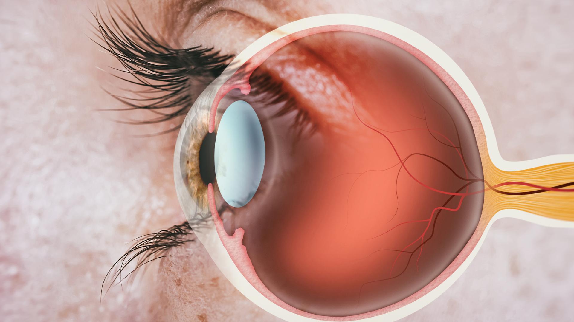
ANDREW SMITH: When you hear the word angle, you should think”drain of the eye.” So, just like a sink has a drain in it, your eye that’s a ball full of water has a drain. Fluid is made inside the eye and has to escape the eye. Why do we call it the angle? We call it the angle because the site of the drainage is right where the iris and the cornea and the sclera meet in the corner of the front part of the eye. You actually can’t visualize it unless you put a lens on the eye and use a little mirror to work your way into that angle to see the drainage structures. So anyway, we’ve kind of use the short term “angle” to mean this natural drainage point inside the eye.
JIMMY LIU: Perfect. Thank you for that explanation, Dr. Smith. And can you also further elaborate on the differences in what causes open-angle and closed-angle glaucoma?
ANDREW SMITH: Yes.I think, actually, we should step back a little bit and just make sure we have an understanding that glaucoma is when the optic nerve starts to thin and degenerate, and that is associated with high pressure. And so, in glaucoma, we want to reduce the pressure. The next kind of umbrella term for glaucoma is this open-angle and closed-angle glaucoma. And basically, it just means that you have glaucoma, but your angle or your drainage system is wide open versus you have glaucoma and your drain is being obstructed by something.
And so, it’s easier to conceptualize a closed angle, so let’s start there. What are some of the causes of closed-angle glaucoma? The most common cause is the iris is being pushed forward by the lens of your eye. So, it turns out that the lens of your eye is right behind the iris. And that lens grows over time, and as it grows, it can push the iris forward. And that angle that’s made by the iris and the cornea on the inside part of the eye can become so narrow that it actually starts to touch and scar down. Other things can cause closed angle. You can have a mass behind your eye. A traumatic event that maybe moves the lens forward can do that. There’s actually a lot of different types of causes for closed angle, but they’re all encapsulated by this idea that that angle is being zipped closed by some event.
That’s in contrast with open-angle glaucoma, of which there are a lot of different types of open-angle glaucoma, in which the angle is wide open but the drainage system is likely not functioning. The most common diagnosis of open-angle glaucoma we call primary open-angle glaucoma. In medicine, that word “primary” means we don’t really have a definitive answer. And so, we know that genetics play a role, and there’s other risk factors I think we’ll talk about later, but that’s the most common open-angle glaucoma. But there are others. There’s a glaucoma called pseudoexfoliation, for example. And that’s a glaucoma where the eye is secreting these proteins, and the proteins are plugging the drainage system. But despite that plugging, when you look, the angle is open. It’s not being zipped closed. And so, that’s a type of open-angle glaucoma. There’s another one called pigmentary glaucoma. And it’s the same concept. The pigments of the eye are clogging the drainage system. You might consider an analogy, again, of a sink, and the sink hole is open, but there’s little food particles plugging up the sink, so it’s backing up. Those are different types of open-angle glaucoma. Like I said, there’s a few different types, but the main difference here is that when you look at the drainage system in open-angle glaucoma, it looks open. And in closed-angle glaucoma, it is pinched shut.
JIMMY LIU: Perfect. Thanks so much for that explanation, Dr. Smith, between the differences and causes of open-and closed-angle glaucoma. You mentioned in the previous answer about risk factors for open-angle glaucoma, and you talked about genetics and other things like that. Can you just describe that a little bit further and consider who is most at risk for developing open-angle and closed-angle glaucoma?
ANDREW SMITH: Absolutely.Let’s start with open-angle glaucoma. The main risk factor for open-angle glaucoma is age and family history. Let’s start with age. Usually, open-angle glaucoma starts over the age of 40, although nothing is a “for sure” rule in ophthalmology or in all of medicine. There are definitely cases of open-angle glaucoma before then. But the risk certainly rises as you get older. And then, family history, especially if you have a sibling who has glaucoma, the risk of you getting glaucoma is much higher. Or another scenario I like to ask my patients is, if your mother has glaucoma and your mother has siblings that have glaucoma, then, again, you’re much more likely to have glaucoma, and so you should be evaluated. Other risk factors are ethnicity. People of African descent or Hispanic or Latino ancestry are more likely to develop glaucoma. And then people with cardiovascular disease-the most prominent one being diabetes-they are a little bit more likely to develop open-angle glaucoma. And the last set is people who are very nearsighted have a little bit increased risk of glaucoma as well.
An interesting point about open-angle glaucoma, and really closed-angle glaucoma as well, is we revolve our world around the eye pressure. But if you get into the weeds with a glaucoma specialist, we consider the eye pressure actually a risk factor. And you’ll hear us say that the eye pressure is the only modifiable risk factor. And so, eye pressure can be high and you don’t have glaucoma, meaning that there’s no optic nerve damage happening, the eye pressure is simply high. And so, we look at that as a risk factor for glaucoma, not necessarily that you have or even will develop glaucoma.
And then another interesting finding for glaucoma that’s a risk factor is a thin cornea. That’s in contrast to closed-angle glaucoma. There are some things similar and some things different. Age and family history play an important role in closed-angle glaucoma. But people who have smaller eyes, they tend to be farsighted. And so, if your prescription is something like +2 or +3, if you’re a farsighted person, you’re at higher risk of having closed-angle glaucoma. Additionally, for closed-angle glaucoma, females are a little bit more likely to have closed angle. And then people of Asian ancestry or Inuit ancestry are more likely to develop closed angle, as well. And then lastly, there is something interesting about a risk factor with people who have already narrow angles and they want to close. Certain medications-like medications for allergy, like Benadryl, for instance, or Sudafed-is another one that dilates the pupil a little bit-those are a little bit of a risk factor for increasing the risk of developing closed-angle glaucoma, as well.
And so, unfortunately, you talk about risk factors in any disease, and glaucoma is no exception. It’s always a little bit difficult to pin those exactly to the diagnosis. But if you have one of these things that we mentioned, it’s a good idea to go ahead and get screened and follow up with an eye provider to make sure that they are watching for glaucoma. As we know, and I know we didn’t get into this at the beginning, but glaucoma is very silent. Most people don’t notice they have it until the very end stages of the disease, and so monitoring, as well as screening, plays a very important role.
JIMMY LIU: Perfect. Thanks so much for that comprehensive explanation, Dr. Smith.So going toward the symptom side of both open- and closed-angle glaucoma, can you describe the differences in the symptoms between the two?
ANDREW SMITH: Yes.Generally, like I said, glaucoma doesn’t have a lot of symptoms until late into the disease. It tends to destroy the peripheral vision first, and it works its way inside. There is an exception to that, and that is usually in some type of closed-angle glaucoma. And so, we’ll talk about that now. So if your angle, your drainage system closes very suddenly, we call that event an acute angle closure crisis. And if you are in crisis, you tend to be symptomatic. So the symptoms you experience are severe eye pain, the eye often turns red, the vision becomes blurry, and people often say they see halos around lights. Often, there is pain also in the head, so you have some type of headaches. Most commonly in patients, I’ve noticed, they form a headache right above the eyebrow. Right along the eyebrow is a very common place to feel that headache. And then in addition, nausea is very common thing. Vomiting can happen. And so, it’s usually pretty dramatic for people. Most people will seek care, but if those things happen to you, you want to go to the emergency room because that is an emergency. The glaucoma is progressing extremely rapidly, and it leads to total blindness if it’s not managed in an appropriate, timely fashion.
There are open-angle glaucomas that can get the pressure up pretty high, but typically this is not happening. Mostly, open-angle glaucoma is a very slow disease without symptoms. It takes out the peripheral vision first, like I said, and then makes its way ahead. And so, it’s really sad. Most people don’t notice until it’s really far into their peripheral vision. And unfortunately, once you incur nerve damage from glaucoma, we don’t have ways to regenerate the optic nerve, and so that vision is lost. And again, highlighting this idea I think we heard at the beginning of glaucoma being called “the silent thief of sight.” And again, highlighting the need to screen people for glaucoma and make sure that we’re watching glaucoma carefully if we have a high suspicion of it.
JIMMY LIU: Thanks so much, Dr. Smith. So,moving on from the symptoms to diagnosis: What does the diagnostic process look like? And what tests help distinguish between open-angle and closed-angle glaucoma?
ANDREW SMITH: Soto understand the diagnosis, I just want to repeat that glaucoma is when the optic nerve is degenerating. And so, the diagnostic tests revolve around looking at that optic nerve and seeing how it functions. So, how we do that in clinic is we run a couple of tests. One is called OCT or optical coherence tomography. That’s just a quick picture of the eye. And you can think of it as if measuring a thickness of your optic nerve. And it compares that thickness to people of your same age, so age-matched controls. And it will flag it if it’s, “Hey, you’re a little bit thinner than someone of your same age,” or even flag it if it’s a little bit thicker, which would be a whole different ballgame to play. And then the other thing with that test is you can follow that test, that thickness over time. You shoot these pictures over time and see if the glaucoma is progressing or how fast it is progressing.
Another test we’ll do in clinic is called a visual field test. Like we talked about, the center vision is typically not affected in glaucoma. And so, just simply reading the chart is not a way to distinguish glaucoma versus non-glaucoma. A visual field test tests your peripheral vision. Typically, you sit in a machine, you cover one eye, you’re given a little clicker in one hand, and then you stare straight ahead at a light, and then other lights in the machine will flicker. And every time you see a light flicker, you’re supposed to click the button. And this allows the test to kind of formulate how far your peripheral vision goes out. And there are certain patterns of visual field loss that are typical of glaucoma, and it will map that out for the physician, who can then correlate it with the other exam findings and see if there’s glaucoma going on.
The next important part of the workup is an exam, and it starts with an eye pressure, so we’ll measure pressure. One key thing about eye pressure is just because you have high pressure does not mean that you necessarily have glaucoma. Like we talked about before, we look at it as a risk factor. And there are plenty of people with quote-unquote “high pressure” or higher-than-the-average-population pressure, and they actually don’t ever develop glaucoma. And on the flip side, there are people whose pressure is quite normal, but their optic nerve is thinning and degenerating in a glaucoma pattern. And so, those people you still treat the same way. You lower the eye pressure from its starting point.
But eye pressure is simply a risk factor; it doesn’t necessarily diagnose glaucoma. After you measure the eye pressure, you look at the eye generally. There are certain findings of different types of glaucoma. We talked about, for instance, pigmentary glaucoma or pseudoexfoliation. You can look for those findings on the exam, and then you also look at the optic nerve itself. In glaucoma, the optic nerve takes on a certain shape, and we call that shape cupping. And it’s a little hard to explain over a phone call without an image, but basically, the optic nerve, as it loses its nerve fibers, the center part starts to become a little bit larger and go back. And there are other things about the blood vessels, and maybe even a little bit of bleeding on the nerve, that you can look at to tell if there is glaucoma happening.
And then the last part of the exam I’ll mention is gonioscopy. And the reason that’s important is because gonioscopy is what tells you the difference between open-angle and closed-angle glaucoma. Gonioscopy refers to someone sitting in the exam chair at the slit lamp, which is that microscope that we use, and you give the patient a numbing drop on the eye, and you place this lens that you hold onto the eye. It tends to have a mirror on it, and that mirror allows you to see into the angle or the drainage system of the eye. And there are certain structures that are in that drainage system. And if you can see them, then we think of it as open. And if you can’t see them, then we think of it as closed. And that will help direct the treatment based on whether you’re open or closed. And like I said before, it’s important to remember that these open-angle and closed angle-categories are really categories of multiple different types of glaucoma. There’s multiple different types of open and multiple different types of closed, so it just kind of points you in one direction. So, that gonioscopy should be done, I think, at every diagnostic visit. Every new patient visit, every patient should have gonioscopy to make sure that we’re determining which direction to go.
Now, let me say one more thing. I don’t want to take a lot of time here. There is a test, an OCT-type test, that can help determine if your angle is open or closed. And there’s a little bit of research being done on it right now. Most glaucoma specialists will still rely on gonioscopy-it’s cheaper, it’s easier. It’s a tough exam to do. A lot of ophthalmologists will tell you how tough it is to learn to do. But glaucoma specialists, I think, rely more on gonioscopy. I think it’s probably more reliable, although that’s probably being determined, but it’s definitely easier and cheaper, and so a lot of us will do that.
JIMMY LIU: Perfect. Thanks so much, Dr. Smith, for that very comprehensive explanation about the diagnostic process of glaucoma. Andso we moved from symptoms to diagnosis, and now we want to go to treatment: How does treatment differ between open- and closed-angle glaucoma?
ANDREW SMITH: Perfect.Let’s talk about that the treatment is the same for both and the fact that you want to lower the eye pressure. That is currently really the only treatment for glaucoma. As a side note, there’s a lot of research going on about neuroprotection, and that basically means; How do you strengthen the optic nerve so it can withstand more of the pressure or the damage that wants to happen? It’s not really the mainstream treatment at all right now, and it’s still under active research. So, right now, if you visit a glaucoma specialist or an eye provider, we’re going to focus on how to lower the eye pressure. And the main difference, when you look at someone’s angle who’s open and who’s closed, is that if they’re closed, you want to try to halt the closing process. It turns out that most of the eye drops that help to lower the pressure work mostly by turning down how much fluid the eye makes, with a few exceptions. There are some that help the eye flow better, but it works better if the angle is open. So because of that, if you are in a closed-angle situation, we want to either try to open it up or at least halt the process of it wanting to close further.
So, in closed-angle glaucoma, you actually might start with a laser treatment. There’s a laser treatment called a peripheral iridotomy. And what it does is you make a little hole in the iris, or the brown or blue or hazel part of the eye, and that hole in the periphery allows for fluid to come around in a different direction and stop and maybe even open up the drainage system a little bit. We will even do those iridotomies if you don’t have glaucoma quite yet. So, your pressure is normal, your optic nerve is healthy, but if your drainage system is narrowing to the point where it’s almost touching or touching to a certain degree, we will intervene at that stage and say, “Hey, we need to do the iridotomy now to keep it open so you do not develop glaucoma in the future.” And if you’re already developing glaucoma and your pressure is high, we often will start there.
Another way you can open the drainage system is to do cataract surgery. If your cataract is far enough along, instead of doing the iridotomy, then we will just go into the operating room and take out the cataract and try to get you better vision by putting a new lens in the eye. But when you do cataract surgery and take out that lens, then the iris can relax back and we can open the angle that way or, again, at least start to halt the process. So anyway, with closed-angle glaucoma, the first priority really is to try to open the angle to stop the process of it zipping up so that you don’t zip up all the way and the pressure goes up really high.
After that, the treatments are pretty similar. The three categories of treatments for glaucoma are eye drops, laser, and surgery. There is one difference in that there is a laser procedure for open-angle glaucoma only. It is called selective laser trabeculoplasty. And that is not done in closed-angle glaucoma because it requires having an open angle to perform that procedure. Surgery is about the same. You can give someone a new drain in surgery-and these are like tube devices or a surgery called a trabeculectomy-that’s allowing the fluid in the eye to flow in a totally different location.
And then there are these procedures that you can perform on the angle, on the drainage system. You should think of it as, like, enhancing your normal drainage system. Or maybe, again, in analogy of a sink, you could think of it as widening the sinkhole or trying to maybe stick a catheter down the sink and enlarge the pipe kind of thing. And those actually can be done on both. They are more indicated for open-angle glaucoma. In closed-angle glaucoma, you can take the instrument and try to open, like maybe peel the iris away from the drainage system if it is stuck there. And then some of us will try to perform the initial steps of those to try to get the fluid to flow naturally through the natural drainage system. They are easy procedures, which is nice. There is a marketing term called MIGS, basically, or minimally invasive glaucoma surgery. That’s the category these surgeries fall under. They luckily tend to be pretty low risk, pretty simple to do, and quick and easy to recover from, which is nice. They’re not as successful as the bigger surgeries at getting the pressure down, but they’re definitely lower risk, and so a lot of us will start at those, as well. And again, like I said, these MIGS are more for open-angle glaucoma, but they can be attempted in closed-angle glaucoma, as well, if you can open the drainage system enough.
JIMMY LIU: Perfect. Thanks so much, Dr. Smith, for that very comprehensive explanation about the different treatment options for both open-angle andclosed-angle glaucoma. We actually received some listener questions. The first one is just someone would like for you to repeat the test with the name that starts with a G. It’s the last one that you mentioned in the diagnostic section.
ANDREW SMITH: Yes.It’s called gonioscopy.
JIMMY LIU: Perfect. Thank you. And then the second question that a listener hassubmitted, and you talked a little bit about this with the cataract surgery: How risky is cataract surgery after a diagnosis of closed-angle glaucoma? And what about the cataract surgery and open-angle glaucoma and the risk for that, as well?
ANDREW SMITH: Let me first say that people with glaucoma in general are just …when you do cataract surgery, unfortunately, the complications are just a little bit higher. And so, you definitely want to be paying attention and take that surgery seriously. And like I said, that goes for open angle and closed-angle glaucoma. People with closed-angle glaucoma, especially if you are in an acute angle-closure crisis-that’s where it’s kind of an emergency-we will do cataract surgery in that event. And you are more likely to have inflammation after surgery, more likely to have the lens be … let me go back. The lens in your eye is suspended by these little springs. It’s almost like a trampoline. If you picture a trampoline, there’s springs all the way around it in a circle. And that’s how the lens attaches to your eye. And when you are in angle closure crisis, those springs can become weak, and that makes the cataract surgery much more difficult. And it might even make it so that the lens that you want to put inside the eye once you take the cataract out can’t be supported in the traditional fashion and you have to do something else to make sure the lens is suspended in the eye.
And then, additionally, after surgery, with both glaucomas, you can have pressure that is too high, you can have pressure that is too low afterward. You can have inflammation, actually, in the front part of the eye, as well as in the retina-that’s more common in glaucoma. And then, the other thing is, when you do cataract surgery, you’re trying to measure the eye and put in a certain power lens that helps your eye see the best. It has to be tailored to the patient as best we can. And if you don’t have glaucoma, that success rate is a certain percentage, it’s somewhere in the 90s. If you have glaucoma, that definitely falls. So, we call that refractive surprise, meaning that you tried to get the patient at a certain power but it turns out the math formulas that we use are not exactly accurate. So, that’s more common in glaucoma, as well.
So anyway, the long story short, glaucoma, definitely higher complication risk. You definitely want to be paying attention a little bit more. And if you’re in a closed-angle glaucoma situation, you definitely have, I think, a higher risk. Another thing I didn’t cover: You have a higher risk of damage to the front part of the eye. The cornea is more common in closed-angle glaucoma. And that’s because typically the space you have to operate in is quite small. And because it’s so small, it’s harder to move the instruments, and the surgery will cause a little bit more swelling of the cornea, for instance, or even damage to the cornea. And so, unfortunately, that’s just the way it is. But when you’re in an angle closure crisis, certainly, or even when you’re in open-angle glaucoma or closed-angle glaucoma, sometimes the next best step is to take out the cataract and go forward, and so we often do those. The last thing I will say is if you are in closed-angle glaucoma … and not all providers do this, but I often do it. So, you’re already inside the eye. We talked about these angle-based procedures, meaning that you’re treating the drainage system. I almost will always go in and just make sure that the angle opens up all the way by drying down the iris with these instruments that we have. And so if you’re in angle closure, I like to do that.
JIMMY LIU: Thanks so much, Dr. Smith, for that explanation. The last question that we have for you is: How important is adherence to treatment, and what happens when individuals miss either follow-up appointments or miss their doses for their eye drops for their glaucoma?
ANDREW SMITH: I mean, this is a huge question.I thank you for asking it. Adherence to treatment is crucial. But at the same time, we know that people aren’t perfect. I always say eye drops are easier said than done. There are some people who are better at it than others. But if you can take your eye drops on a very consistent basis, it definitely helps. It’s hard for us to know. When you come to clinic and we measure your pressure, say we’re monitoring the glaucoma, that pressure really is just a snapshot in time. And we don’t really know what’s going on with the pressure as much, like in the middle of the night, for instance, or when you’re at home. And if you’re missing doses but you take your drops before coming to clinic, it can lead to kind of misleading pressures for us. We try to pick that up by seeing if the optic nerve is changing despite having low pressures, but it can be difficult. So, taking drops not only helps keep the glaucoma under check, but it also helps us to kind of manage the glaucoma appropriately so we’re not getting too confused in clinic.
Same with missing appointments. Glaucoma, unfortunately, is a disease process that plays out over time; it’s not something that’s static in one spot. And so, because of that, even if things have been going great for 2 years, it’s so important to get checked on a regular basis because that glaucoma can take a left turn and you just want to make sure that you grab that patient and put him back on the road to stabilization. And so, sometimes I feel like the glaucoma patients who especially are stable, you feel the mundane nature of coming to the appointments and everything being okay. But it’s so important to come because you never know when that glaucoma process will play out a little further or maybe play out in a more aggressive manner, and you want to be able to catch that and push it right back down.
And then the last thing to really point out in this situation is the reason adherence to treatment and follow-up is so important is because glaucoma damage is permanent. We can’t get it back. And so, it’s something that’s, of course, stressful, and we need to have a good relationship with your physician. But you need to adhere to treatment best you can and come to the appointments best you can in order to catch the damage if it’s happening so that you can be more aggressive back at it and push things back down the best you can to slow down the process. So, consistency really matters. Again, as physicians, there’s tons of research that shows that adherence to a drop regimen is difficult and is not always feasible for certain types of people. And that’s why we have the surgeries that kind of help us out. But most people with glaucoma are going to need drops, and so if you can take them consistently, it really does help.
JIMMY LIU: Thanks so much, Dr. Smith, for that explanation. We have time for some listener questions. One listener chimed in and asked, “Can you have both types of glaucoma simultaneously, open angle and closed angle?”
ANDREW SMITH: That’s a phenomenal question, actually.The answer is yes. Closed-angle glaucoma, turns out, is a risk factor for open-angle glaucoma. So, let me give you a scenario. Say you have closed-angle glaucoma, and you treat it with the laser peripheral iridotomy, that little laser procedure I was talking about, and then the angle opens up and you’re no longer closed or narrow. Those people actually are at risk for primary open-angle glaucoma, so they can develop a primary open-angle glaucoma after having a closed-angle or narrow-angle situation. So, let’s take the best-case scenario, like, you catch it before. The drainage system is very narrow, it’s starting to zip up, but you catch it before the pressure goes up or before the optic nerve gets damaged. Those patients actually still need to be monitored on a regular basis to make sure they don’t develop other types of glaucoma. So in a way, yes, you can have both scenarios.
JIMMY LIU: Thanks so much, Dr. Smith, for that explanation. The last listener question that we have is they wanted to get your opinion on future treatments for open-and closed-angle glaucoma. You mentioned previously before about neuroprotection, which is super exciting and a really interesting research topic in glaucoma. Is there anything about either neuroprotection or decreasing eye pressure that you are most looking forward to in terms of treatments to glaucoma patients going forward?
ANDREW SMITH: Yeah, definitely.Let’s talk about both. Let me talk about delivery of medications first. There’s been a lot of development in recent years about trying to make these implants, essentially, that will slowly release medication inside your eye-so, you inject the implant into your eye. Those are good right now, but they need to get better. And they will get better, I think. So, it might turn out that instead of relying on someone to take two or three drops two or three times a day, that we end up figuring out a way to inject the medications into the eye to slowly release over time and that we don’t have to worry about putting drops in all the time inside the eye. There are some risks, of course, with doing something like that, but we really are optimistic that that will be the future and will help people with compliance and might even be more effective because also it’s difficult to put eye drops in the eye, let alone the consistency of it. So, I think that will come. There are already two devices on the market that do that. One is only approved for 3 months of use. The other lasts 3 years, it goes actually into the drainage system, and it might become more widespread. But I personally think there are need and room for much improvement on both of those devices, and that will come.
Let’s go to neuroprotection. Neuroprotection is an active area of research. There is a lot of consideration into it. If you ever go and just search things like vitamins or exercises for glaucoma, you will see a myriad of research articles. And to me, I look at it as kind of a mess. So, there’s different vitamins that have been studied that we think might help with optic nerve stability and neuroprotection, and so that is an area of active research right now, and then hopefully, that comes more and more to fruition. There are medications that people think need to be tested, like, instead of a vitamin, more like a medication that will provide neuroprotection. But those, I feel, are-in my reading of the research-earlier in their stages of research, and I don’t see them coming soon, but it is an area of active research.
Another thing is that if someone is progressing at very low pressures-so, their glaucoma is getting worse despite low pressure-a lot of us will look at different types of lifestyle risk factors. For example, some people, their blood pressure is getting too low, and that actually is a little risk factor for progression of glaucoma, that it’s too low. Or maybe another situation is getting migraine headaches or having this phenomenon called Raynaud’s phenomenon. So, there are other things to look at that maybe affect the optic nerve and the nutrition that it gets that already we look at. But in the future, we hope to have more medications or vitamins or diets that hopefully help to strengthen the optic nerve.
JIMMY LIU: Perfect. Thanks so much, Dr. Smith, for giving all of our listenerssome hopeful look into all the different treatments for glaucoma into the future. That’s all the time we have today for questions. Thank you so much again, Dr. Smith, for answering so many of our questions and all the information you shared with us. To our listeners, thank you so much for joining our Glaucoma Chat. I sincerely hope you found it helpful. I would like to mention that BrightFocus Foundation and the American Glaucoma Society have websites with a wealth of information about glaucoma. Please visit www.BrightFocus.org and www.AmericanGlaucomaSociety.net to learn more. And Dr. Smith, before we close, is there one message you hope individuals take away from understanding these two types of glaucoma?
ANDREW SMITH: Yes. A lot of patients come in after being diagnosed with glaucoma and understandably are very scared about what is going to happen with their vision. But I think if I had to say one messageit’s that glaucoma is manageable. I often use this phrase with my patients: “Glaucoma is a lot of work, and we’re going to have to work at it.” But we can often be very successful in preserving the vision, and so I think there is optimism to be had in facing glaucoma, and I think patients should share in that optimism. And we’ve definitely gotten better at taking care of glaucoma and preventing blindness. And there is a big push to have more devices and more treatments available to patients and that the field will continue to expand and to get better. So, I’m optimistic about the treatments. When I talk to patients, I hope to share that optimism and to feel like the glaucoma can be managed or can be slowed down, and I think that’s good. I also want to just say thank you so much for having me on, and it’s really an honor to just be in front of people talking about glaucoma. It’s one of my favorite things to do, so thank you so much for the opportunity.
JIMMY LIU: Awesome. And thank you so much, Dr. Smith, for taking time out of your busy schedule to come speak with us and everyone on this call about glaucoma. We atBrightFocus are also very passionate about glaucoma and eventually curing this disease that is so prevalent in our society. So, thank you so much again for that advice. Our next Glaucoma Chat will be on Wednesday, March 11, 2026. Thanks again for joining us, and this concludes today’s Glaucoma Chat.
Useful Resources and Key Terms
BrightFocus Foundation: (800) 437-2423 or visit us at www.BrightFocus.org. Available resources include-
Helpful treatment options or resources mentioned during the Chat include-
- optical coherence tomography (OCT)
- gonioscopy
- selective laser trabeculoplasty
- peripheral iridotomy
- minimally invasive glaucoma surgery (MIGS)