


Our Approach to Glaucoma Research
BrightFocus takes a 360-degree approach to funding innovative scientific research worldwide to defeat glaucoma. We explore the full range of scientific paths toward better treatments and, ultimately, a cure. By investing in a wide range of innovative scientific approaches, we leave no stone unturned in the quest for a cure.
Controlling Eye Pressure in New Ways
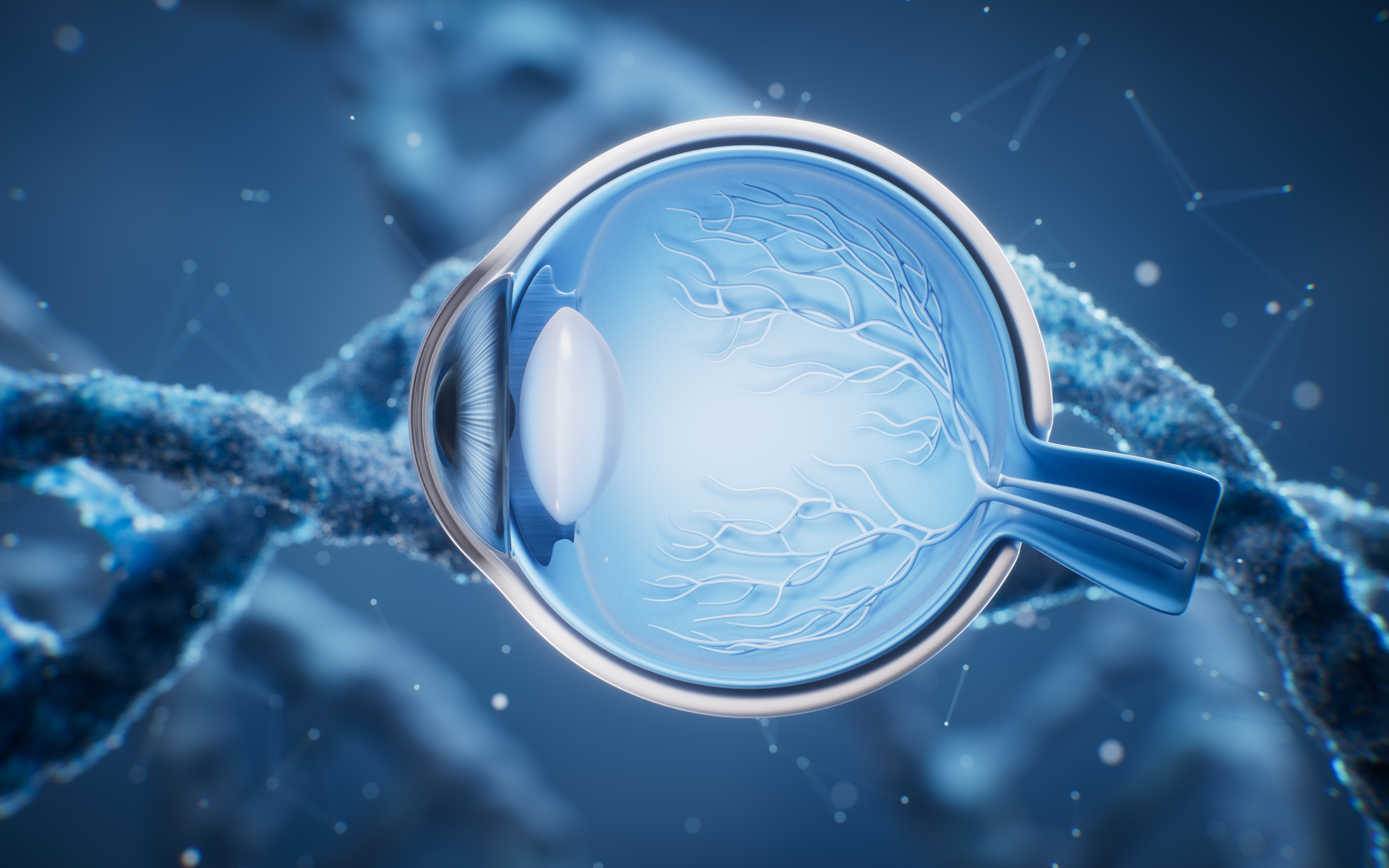
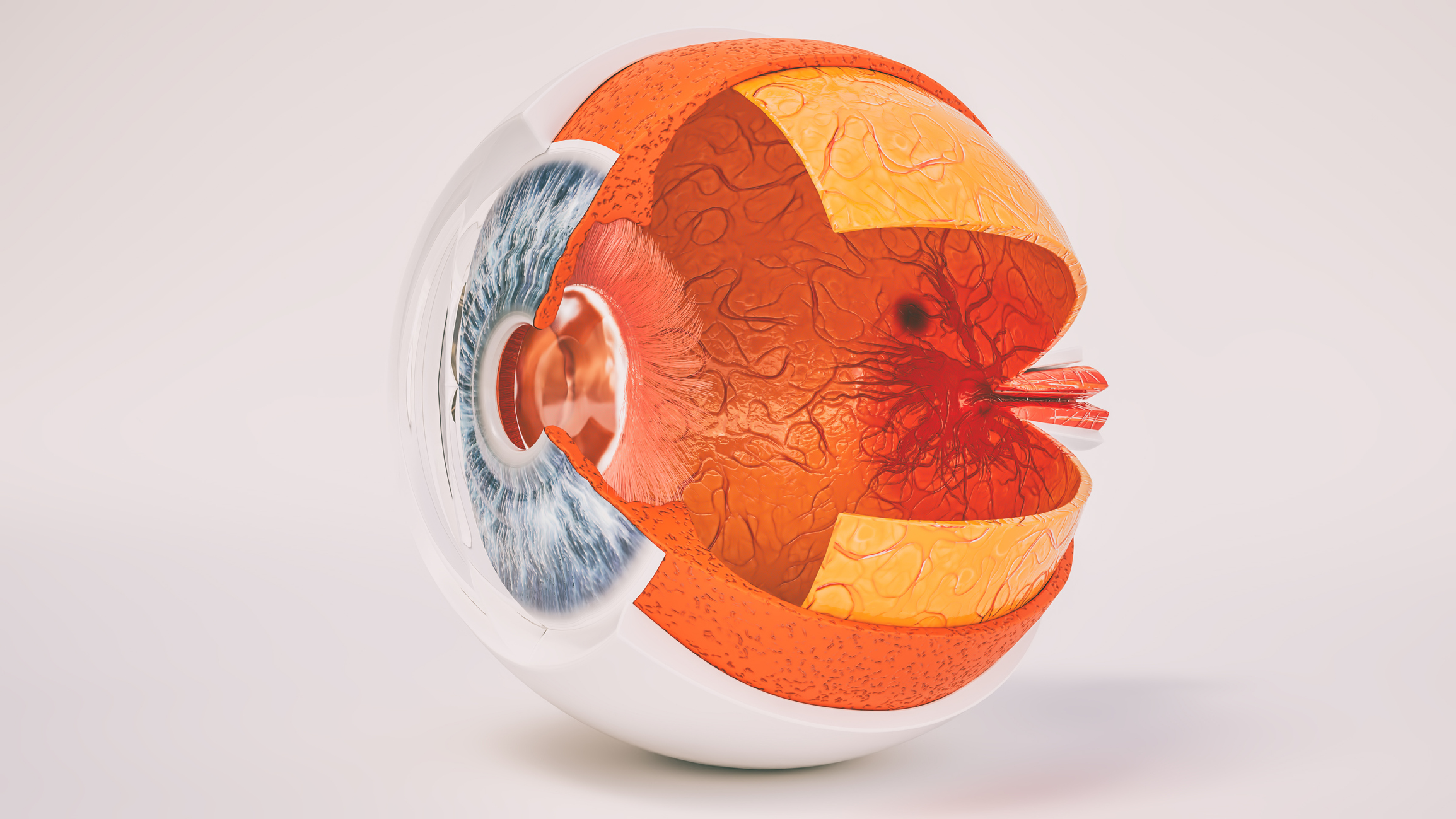
Elevated eye pressure, or intraocular pressure (IOP), is present in most forms of glaucoma when aqueous humor, the fluid that constantly bathes the front of the eye, cannot drain properly. Normally, the fluid drains through a spongy tissue known as the trabecular meshwork and flows into Schlemm’s canal, a ring-like passageway that then delivers it to the bloodstream. Blockages and other forms of resistance to aqueous humor outflow can raise eye pressure. Other factors are fluid volume and trabecular meshwork stiffness, which is reported to be 20 times higher in glaucoma than in healthy eyes. With critical National Glaucoma Research funding, grantees are unraveling novel mechanisms that regulate eye pressure and are exploring new ways to decrease stiffness and control eye pressure.
Understanding What Causes Glaucoma
Ultimately, glaucoma threatens sight by damaging the optic nerve at the back of the eye, which carries light signals from the eye to the brain. Our knowledge of how and when glaucoma damages nerve cells remains imprecise. It’s linked mostly to chronic pressure increases inside the eye, referred to as elevated intraocular pressure (IOP), which may arise from the eye’s inability to drain fluid properly. National Glaucoma Research is funding studies on genetics, including studies addressing racial and ethnic disparities in disease incidence and onset. Other projects include developing more sensitive methods for studying onset and projects to develop new research models to promote a better understanding of glaucoma that may lead to new therapies.
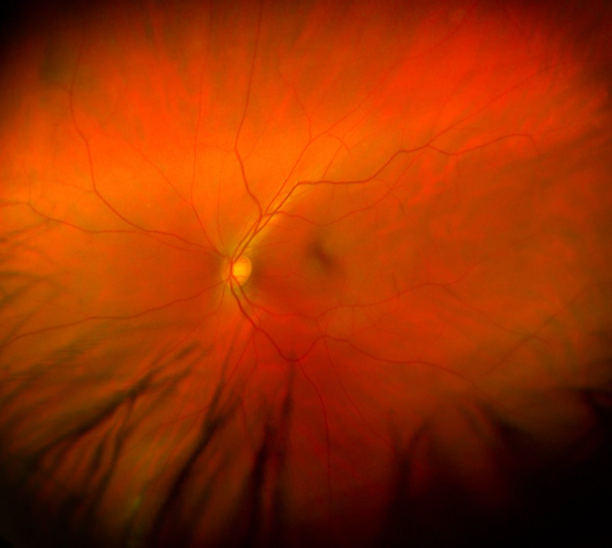
Imaging & Exploring the Eye-Brain Connection
Eye changes associated with glaucoma contribute to tiny blind spots, known as visual field defects, which can advance to vision loss and blindness. The speed and likelihood of this progression vary from person to person. Early diagnosis is key, and considerable progress has been made in eye imaging to detect the tiniest changes preceding glaucoma. National Glaucoma Research grantees are developing and leveraging new technologies to look at individual retinal ganglion cells (RGCs) of the eye and their nerve fibers, which carry light signals to the brain—a challenging task because RGCs are almost transparent and difficult to image. Scientists also are investigating disruptions in how cells communicate in glaucoma. The findings could result in earlier detection of and new ways to treat glaucoma.
Predicting Outcomes & Other Treatment Innovations
Approved treatments for glaucoma primarily focus on lowering eye pressure. Many therapies involving eye drops or surgery lower eye pressure effectively, but most require skill and consistency to achieve results or, as with surgery, present recognizable risks.
More reliable treatments and new therapies to address the underlying causes of glaucoma beyond changes in intraocular pressure are needed. National Glaucoma Research grantees are working to develop drugs that will lower eye pressure and protect against nerve cell injury and death, as well as genome-editing approaches to restore the function of trabecular meshwork (a spongy tissue that drains fluids from the eye). Additional therapies include advancing stem cell transplantation, promoting lifestyle interventions, and identifying strategies to communicate genetic testing with at-risk individuals.
Protecting & Regenerating the Optic Nerve
Unlike most cells in the body, which repair themselves, the nerve cells providing our vision don’t regrow once damaged. National Glaucoma Research is supporting research into ways of protecting cells threatened by advancing glaucoma and regenerating those cells after vision loss. The focus of these efforts is to replace and reconnect retinal ganglion cells (RGCs), the nerve cells that make up the optic nerve and carry visual signals over axons, long threadlike tails extending from the eye to the brain. This is a sophisticated undertaking, given how RGCs are wired into the brain. Another focus is to develop neuroprotective drugs and therapies that will help nourish and support fragile RGCs to ensure their long-term viability.
News
Explore Our Research
Understanding the Causes of Steroid-Induced Glaucoma
A commonly used medication is linked to increased risk of glaucoma. A BrightFocus National Glaucoma Research-funded scientist is trying to understand why.
Article
Focusing on the Future: Key Takeaways from the 2025 Glaucoma Fast Track
BrightFocus Foundation’s 2025 Glaucoma Fast Track brought together early-career and established scientists to provide a thorough overview of the biology, diagnosis, and treatment of glaucoma.

Article
How Early Research Funding Is Transforming Glaucoma Detection
If you’ve been diagnosed with glaucoma in the past 15 years, you have likely unknowingly witnessed what early research funding can do for the 4 million Americans living with this sight-stealing disease.
Article
Protecting Vision: A New Approach to Scar-Free Healing From Glaucoma Surgery
BrightFocus National Glaucoma Research-funded scientist Jennifer Fan Gaskin, MD, is exploring safer, more effective treatments to prevent scarring after glaucoma filtration surgery, also known as trabeculectomy.
Article
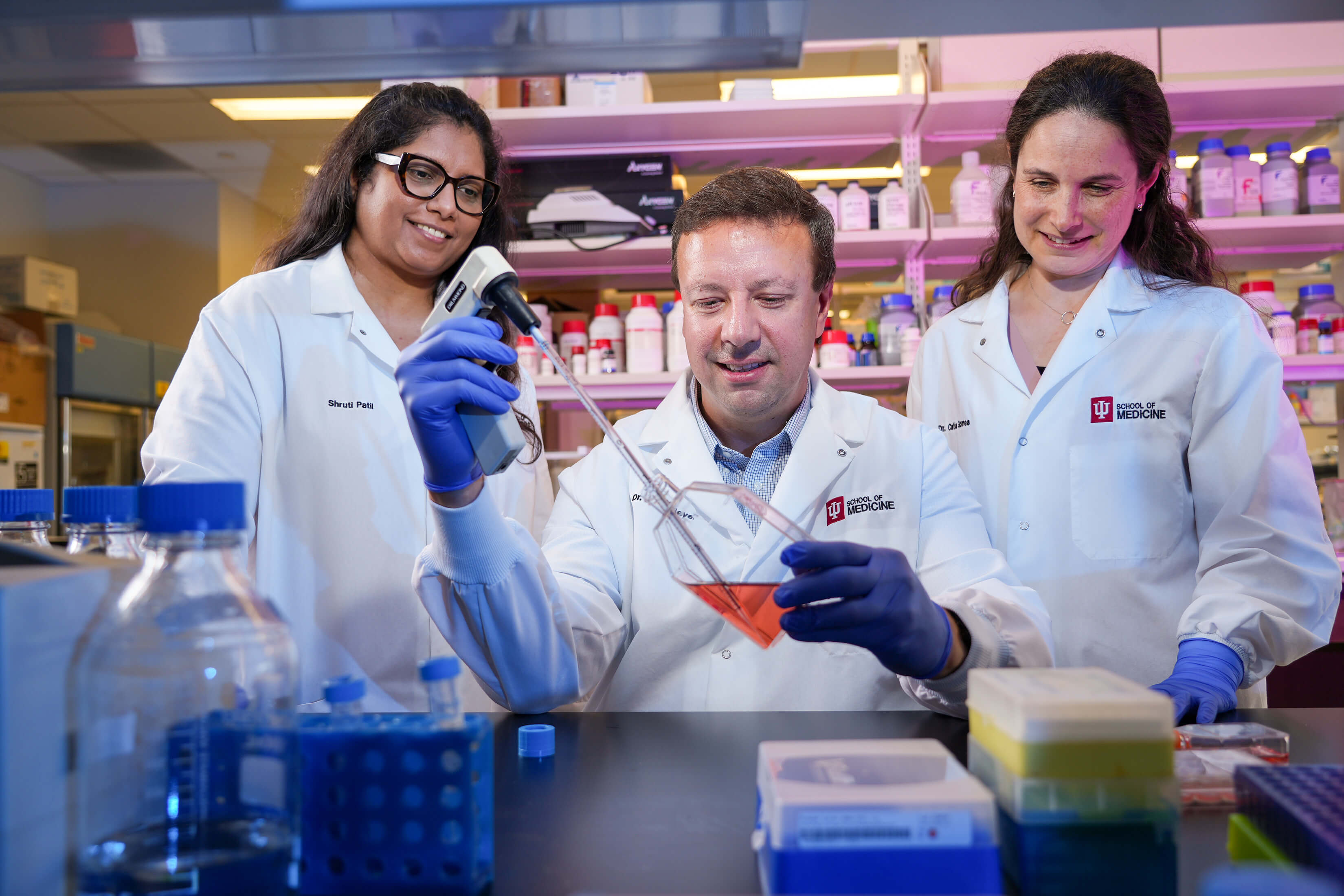
National Glaucoma Research Grantee Receives $2.2 Million Follow-On Award Aimed at Curing Blindness
Additional funding is propelling BrightFocus-funded scientist Jason Meyer, PhD, toward advances in whole-eye transplantation.

Article
Harnessing Artificial Intelligence to Transform Glaucoma Detection
A National Glaucoma Research-funded scientist is pioneering the use of artificial intelligence and telemedicine to detect glaucoma earlier, improve access to care, and prevent irreversible blindness.

Article
How Some Eye Cells Survive Glaucoma
BrightFocus National Glaucoma Research-funded scientist Mengya Zhao, PhD, uses a unique combination of techniques to understand why certain retinal cells stay strong in glaucoma while others don’t.
Article
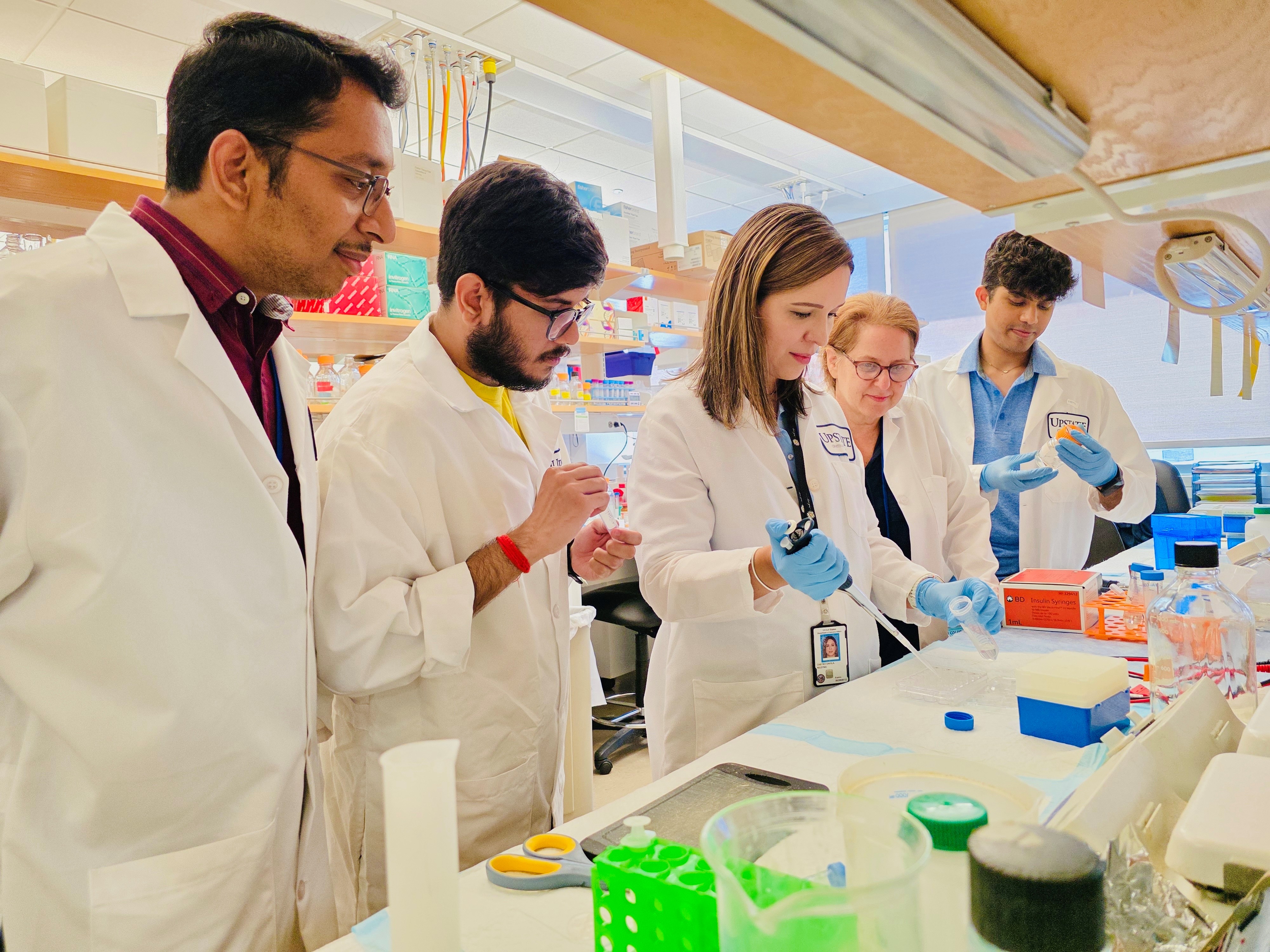
A Key Model to Study Cellular Functions to Better Treat Glaucoma
BrightFocus National Glaucoma Research-funded scientist Samuel Herberg, PhD, is using a 3D model of the fluid drainage tissue that could improve our ability to understand and treat glaucoma.
Article
How the Breakdown of Cellular ‘Powerhouses’ Drives Exfoliation Glaucoma
A study yields new insights that could inspire novel treatments for exfoliation glaucoma.