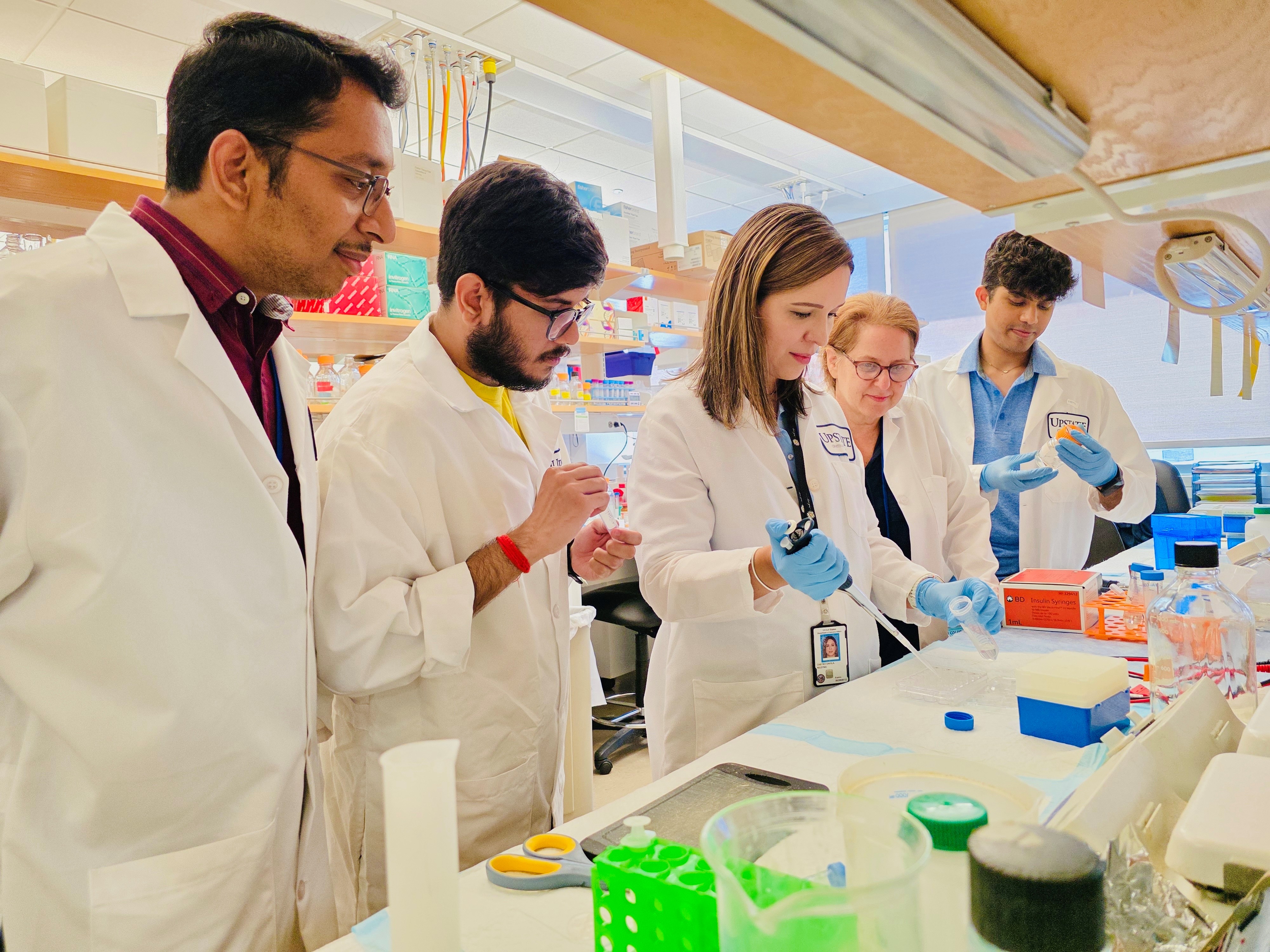
The Bernstein research team at SUNY Upstate Medical University
Key Takeaways
A study led by Arunkumar Venkatesan, PhD, and Audrey Bernstein, PhD, links defects in the mitochondria of eye cells to the development of exfoliation glaucoma.
These dysfunctional mitochondria may contribute to the inability of the cells in the eye to clear misfolded proteins, leading to the accumulation of white, flaky substances that block fluid drainage in the eye, increasing pressure and causing vision loss.
The study illustrated how two drugs could reverse mitochondrial abnormalities in an exfoliation glaucoma model, promoting the formation of healthy mitochondria. This suggests that restoring mitochondrial function may be a promising strategy for treating XFG and potentially preventing vision loss.
Exfoliation glaucoma (XFG; also known as pseudoexfoliation glaucoma) is a more severe form of open-angle glaucoma and a blinding disease. It is characterized by a clumpy, white, flaky substance that blocks fluids from draining from the eye. This increases eye pressure, ultimately leading to vision loss. XFG, for which there is no treatment, affects an estimated 25% of the 60 million cases of open-angle glaucoma worldwide. But what causes these deposits to form in the first place?
A new first-in-class study demonstrates that XFG is characterized by defects in the mitochondria of eye cells. Mitochondria are the cellular powerhouses that give the cell energy to carry out almost every function, including breaking down and clearing misfolded proteins that form in people with XFG. The researchers believe they’ve accomplished a milestone by repurposing a drug that clears the damaged mitochondria to improve cell health in a glaucoma model.
The research out of SUNY Upstate Medical University was led by Arunkumar Venkatesan, PhD, a recipient of joint funding from BrightFocus Foundation and The Glaucoma Foundation, alongside corresponding author Audrey Bernstein, PhD, a previous BrightFocus National Glaucoma Research grant recipient. Dr. Venkatesan is probing how misfolded proteins contribute to XFG, and how mitochondrial dysfunction leads to cell damage that ultimately causes vision loss. He hopes his findings will inspire new methods for clearing exfoliation material before it forms in the eye and for restoring mitochondrial function.
The Role of Misfolded Proteins
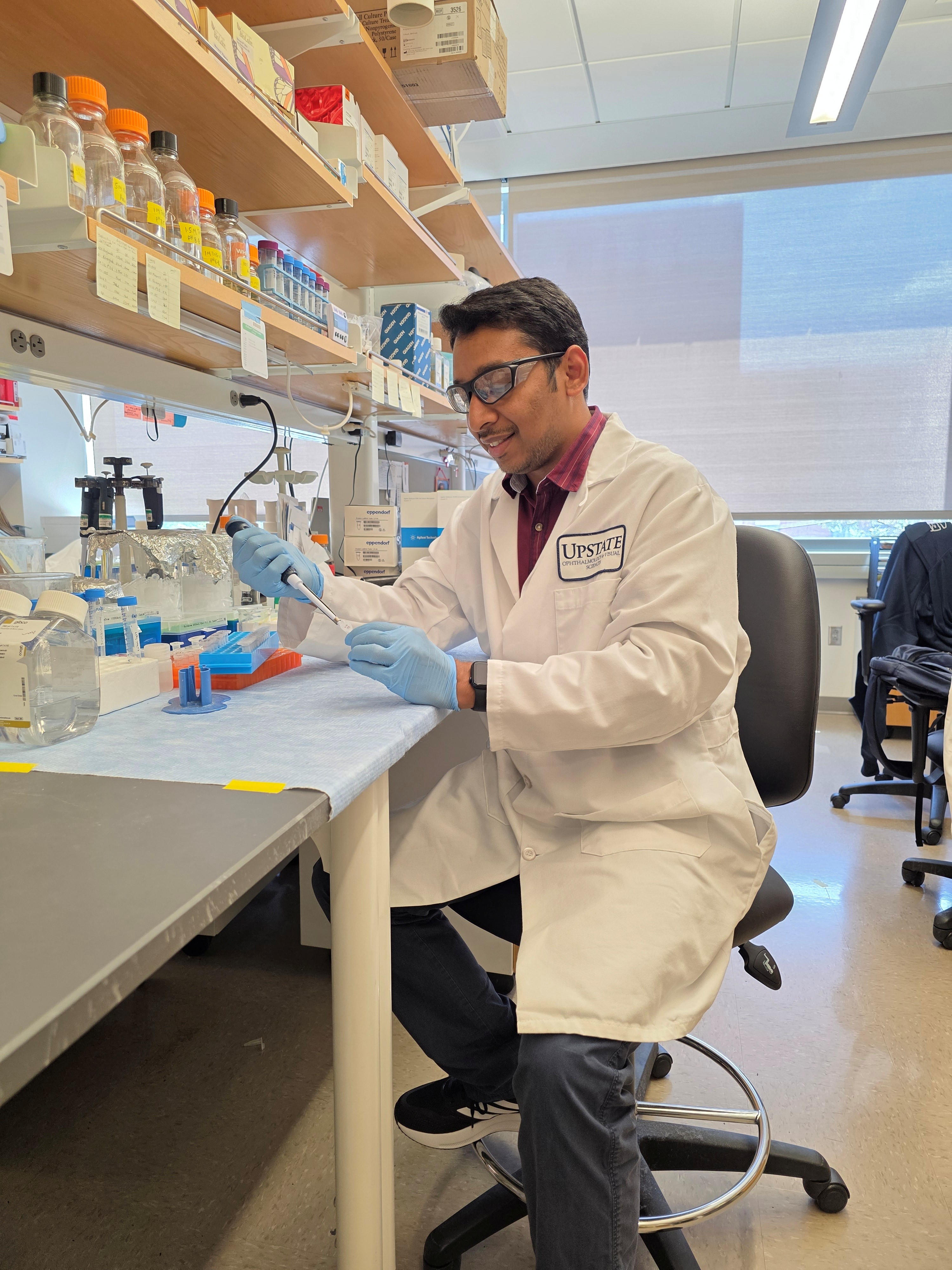
Arunkumar Venkatesan, PhD, BrightFocus Foundation grant recipient
Dr. Venkatesan’s focus on mitochondria stems from his understanding of what drives healthy vision. “Our eyes are amazing organs that work nonstop to capture and send visual information to the brain, a process that requires a tremendous amount of energy. Mitochondria produce energy in the form of ATP (adenosine triphosphate) to fuel these demanding tasks,” Dr. Venkatesan said.
For the new paper published in Free Radical Biology and Medicine, Dr. Venkatesan and his colleagues used a cellular model of XFG to investigate the role of mitochondrial abnormalities in the disease. They further linked this mitochondrial dysfunction to unstable microtubules—scaffolds that preserve the structural integrity of cells. They went on to test drugs in the XFG model to investigate their effect on mitochondria.
Dr. Venkatesan believes that XFG is similar to other age-related diseases that are driven by a build-up of misfolded proteins. These diseases are also marked by the breakdown of processes that would normally destroy abnormal protein aggregates. The presence of failing mitochondria in eye cells could also lead to a condition known as oxidative stress, he said, “putting the cells into a loop of stress and decline” that may drive the progression of XFG. Defects in protein misfolding, mitochondrial dysfunction, and oxidative stress are common players in various types of open-angle glaucoma, as discussed in their recent review paper published in Frontiers in Cell and Developmental Biology, Dr. Venkatesan added.
“To keep vision sharp and eye tissue healthy, especially the retina and optic nerve, cells need a steady and abundant supply of ATP,” Dr. Venkatesan added. “Without this, the health of ocular cells can be compromised, and the cells can become damaged, ultimately leading to vision loss.”
Finding Potential Drug Therapies
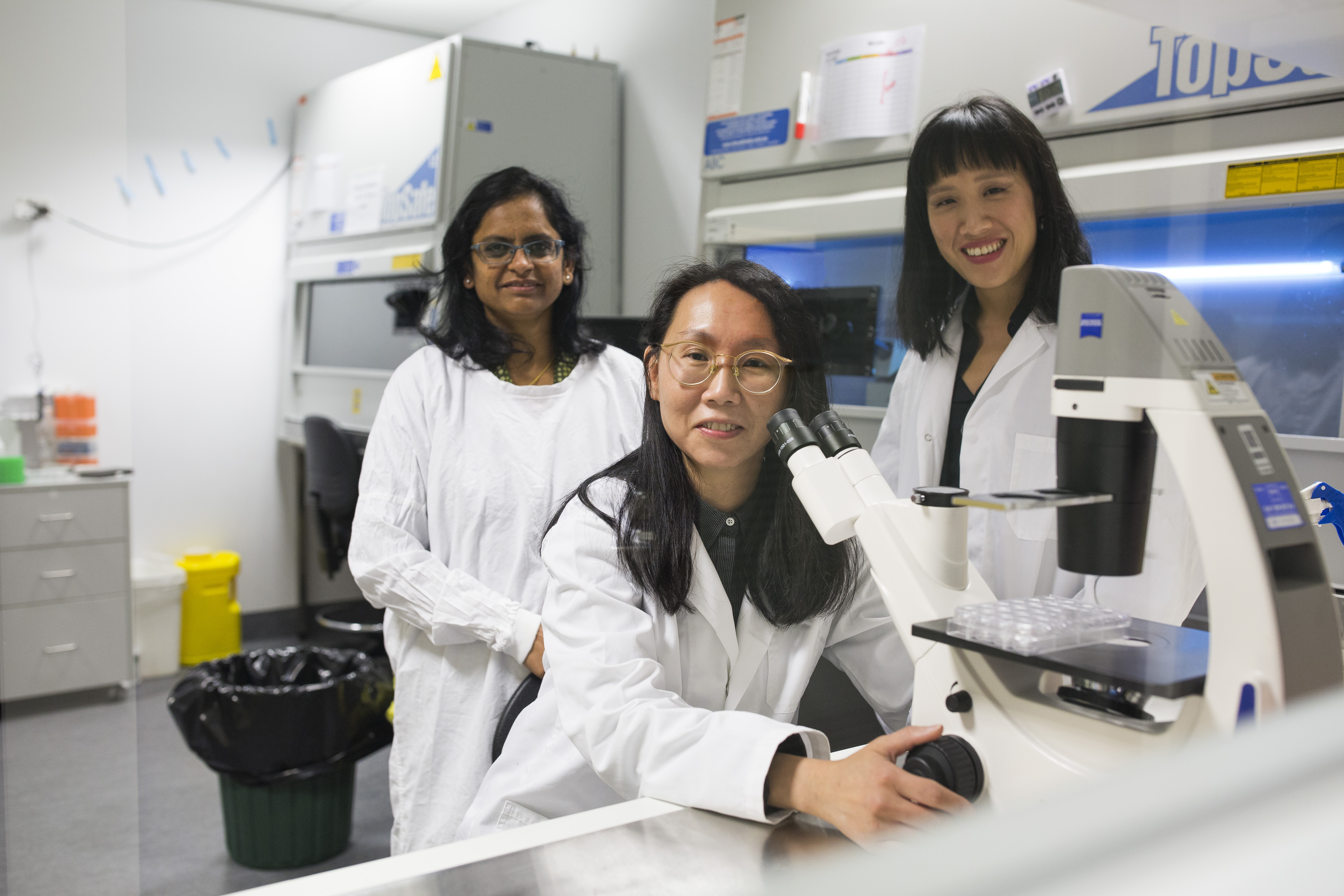
Audrey Bernstein, PhD, National Glaucoma Research grant recipient
During the study, Dr. Venkatesan’s team reversed mitochondrial abnormalities in the XFG model with two drugs previously shown to have positive effects on mitochondrial health: Urolithin A, a natural compound, and nicotinamide ribose, a type of vitamin B3. The drugs worked by clearing the unhealthy mitochondria, opening the door to the formation of healthy, functional mitochondria.
The positive effects of the drugs on XFG will need to be further confirmed in lab models before they can be tested in people. But Dr. Venkatesan is optimistic that his research could contribute to the development of innovative treatments for this type of glaucoma. “This promising finding opens the door for more research into the possibility of restoring mitochondria to help address [protein] defects and clear away the exfoliation material before it forms in the eye,” he said.
Future Directions
Dr. Venkatesan has several additional studies underway that he hopes will build upon the insights he has gained into XFG. Alongside working on novel therapeutic targets to improve mitochondrial function, he and his colleagues are investigating whether there are other cell biological defects in XFG, such as defects in the cellular degradation system that may contribute to producing the disease pathology. They are also delving deeper into the protein-folding defects they observed to better understand how drainage-blocking flaky clumps develop in eye cells.
“Our goal is to use this foundational research to identify a therapy to slow or reverse XFG disease in patients,” he said.
He credits the joint funding for its “major impact” on his XFG research. “We are grateful for the support,” he said. “The work couldn’t have been done without this award.”
Tuteja S, Zeppieri M, Chawla H. Pseudoexfoliation syndrome and glaucoma. In: StatPearls [Internet]. Treasure Island (FL): StatPearls Publishing; 2025 Jan–. Updated 2023 May 31.
BrightFocus Foundation is a premier global nonprofit funder of research to defeat Alzheimer’s, macular degeneration, and glaucoma. Since its inception more than 50 years ago, BrightFocus and its flagship research programs—Alzheimer’s Disease Research, Macular Degeneration Research, and National Glaucoma Research—has awarded more than $330 million in research grants to scientists around the world, catalyzing thousands of scientific breakthroughs, life-enhancing treatments, and diagnostic tools. We also share the latest research findings, expert information, and resources to empower the millions impacted by these devastating diseases. Learn more at brightfocus.org.
Disclaimer: The information provided here is a public service of BrightFocus Foundation and is not intended to constitute medical advice. Please consult your physician for personalized medical, dietary, and/or exercise advice. Any medications or supplements should only be taken under medical supervision. BrightFocus Foundation does not endorse any medical products or therapies.
Protecting Vision: A New Approach to Scar-Free Healing From Glaucoma Surgery
BrightFocus National Glaucoma Research-funded scientist Jennifer Fan Gaskin, MD, is exploring safer, more effective treatments to prevent scarring after glaucoma filtration surgery, also known as trabeculectomy.
Article
National Glaucoma Research Grantee Receives $2.2 Million Follow-On Award Aimed at Curing Blindness
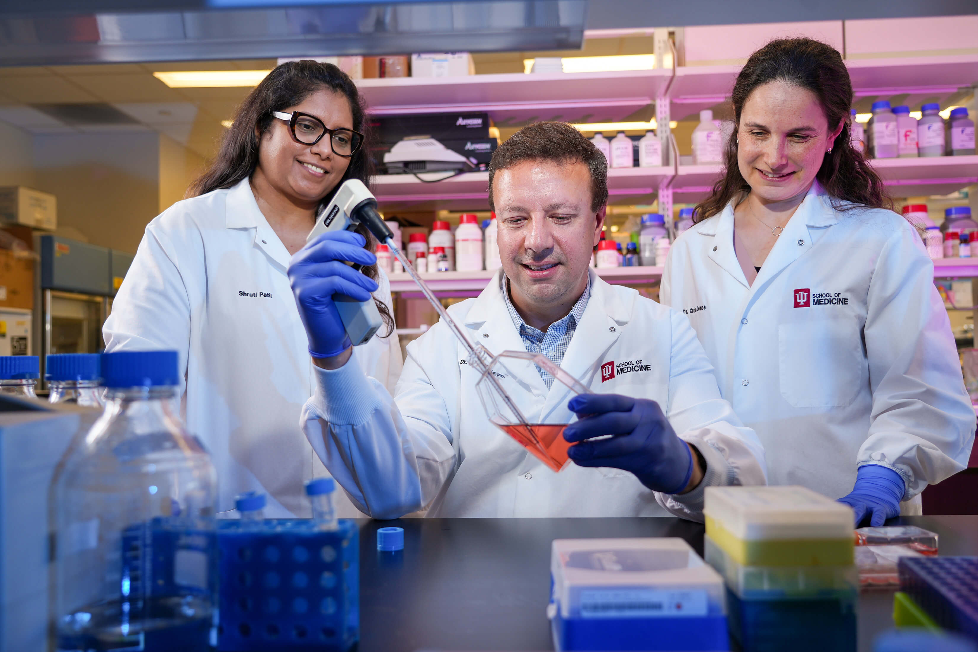
Additional funding is propelling BrightFocus-funded scientist Jason Meyer, PhD, toward advances in whole-eye transplantation.
Article
Harnessing Artificial Intelligence to Transform Glaucoma Detection
A National Glaucoma Research-funded scientist is pioneering the use of artificial intelligence and telemedicine to detect glaucoma earlier, improve access to care, and prevent irreversible blindness.
Article
How Some Eye Cells Survive Glaucoma
BrightFocus National Glaucoma Research-funded scientist Mengya Zhao, PhD, uses a unique combination of techniques to understand why certain retinal cells stay strong in glaucoma while others don’t.
Article
A Key Model to Study Cellular Functions to Better Treat Glaucoma
BrightFocus National Glaucoma Research-funded scientist Samuel Herberg, PhD, is using a 3D model of the fluid drainage tissue that could improve our ability to understand and treat glaucoma.
Article
Looking Beyond Pressure-Lowering Drugs to Treat Glaucoma
A National Glaucoma Research-funded scientist is building a 3D model of the optic nerve that she hopes will solve unanswered questions about glaucoma—and inspire new treatments.
Your support helps fund critical research that could prevent vision loss, provide valuable information to the public, and cure this sight-stealing disease.