Dr. Dominik Paquet, a BrightFocus Alzheimer's Disease Research grant recipient, has developed new, groundbreaking models using lab-grown human brain tissue that better mimic Alzheimer's disease.
While previous models have taught us a lot about Alzheimer's, animals and humans process key proteins differently, and these differences can affect how well potential treatments work.
These models could give researchers a faster, more accurate way to test new Alzheimer's treatments before they reach clinical trials.
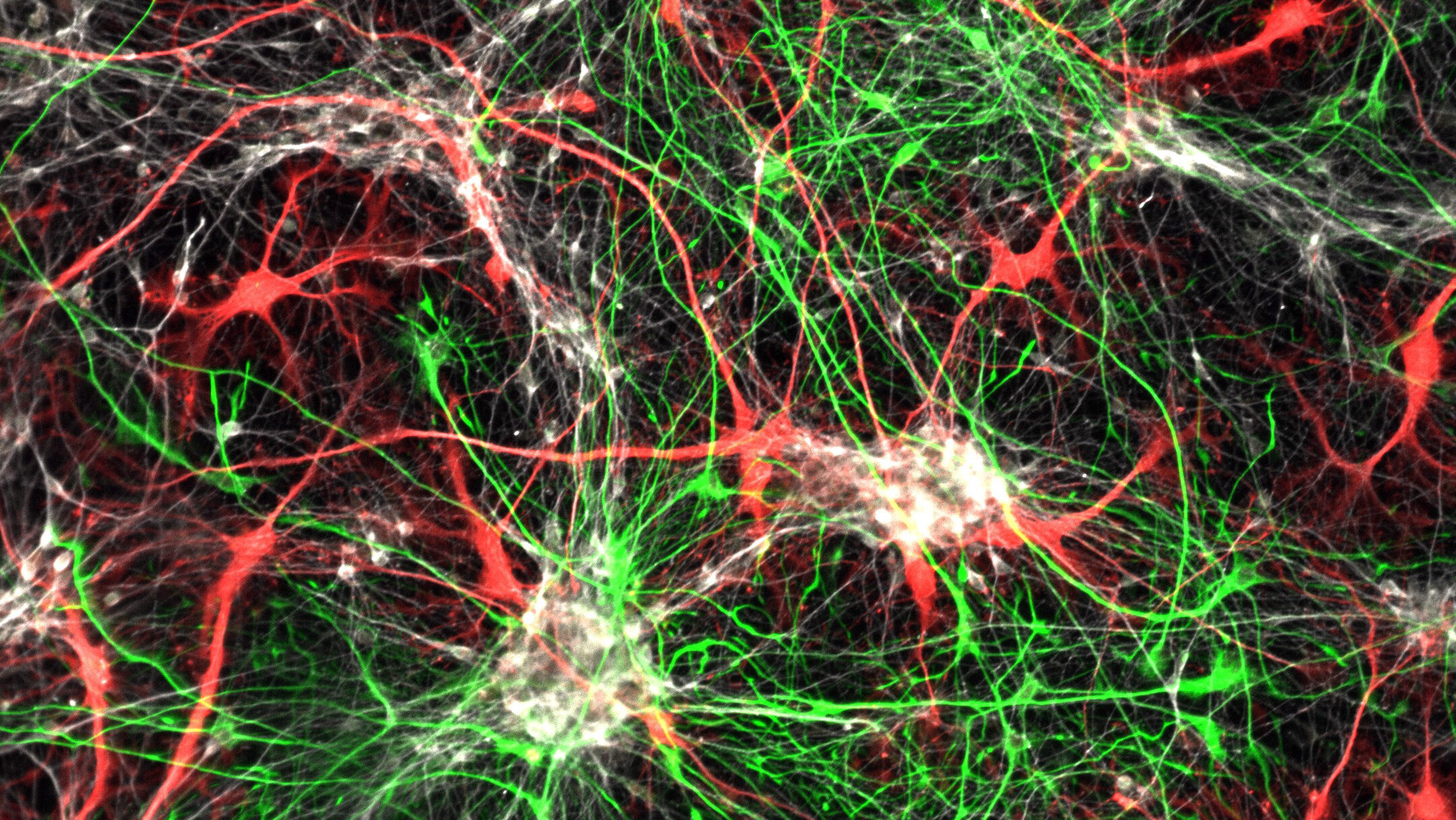
Long-term co-culture of iPSC-derived human cortical neurons and astrocytes stained for neurons in white and glia in red and green. Image courtesy of Dr. Paquet
For decades, studies in animals have taught us much of what we know about Alzheimer’s disease. To take the next big step forward, BrightFocus Alzheimer’s Disease Research grant recipient Dominik Paquet, PhD, thinks the path ahead lies in models built around human brains.
His team developed new, groundbreaking models using lab-grown human brain tissue, aiming to capture disease mechanisms that can’t be studied in animal models alone. The goal is to build a human-centered platform on which to test Alzheimer’s therapies.
Why human-centered models matter
Animal models have been essential for understanding how the brain functions, and how diseases like Alzheimer’s disrupt it. But they have their shortcomings.
One of those key differences involves tau, a protein that keeps nerve cells structurally sound. In Alzheimer’s disease, tau clumps together, and this buildup is tied to cell death. Animals and humans regulate tau differently, and that gap has complicated translating mouse findings into working drugs. Dr. Paquet’s team believes the connection between two important proteins that contribute to the development of Alzheimer’s—amyloid plaques and tau—may have a human-specific piece that only models based on human cells can reveal.
“This is an area where the animal models are not that helpful and where the human brain tissue models can make a difference,” Dr. Paquet explained.
Dr. Paquet’s lab is building models of human brain tissue that closely mirror the cell types of real human brains. These include neurons, cells that fire electrical signals to communicate with one another, and glia, which support neurons and regulate immune response.
Building human brain tissue
But building those models wasn’t easy. The stem cells they use have to be exactly the right age: too young, and they become the wrong kind of cell; too old, and they stop forming tissue altogether. The model also relies on multiple cell types that mature on different timelines yet must grow together in sync, a balance that took years of trial and error to find.
Once the tissue was working, the team faced a second challenge: giving it Alzheimer’s disease. That meant editing the cells’ DNA using a precise gene-editing tool called CRISPR, with each edit made in careful sequence.
“Imagine that our genome is a library containing 3 billion letters (which is about 33,000 books),” Dr. Paquet said. “We just want to change one letter in one book, but need to be sure that we haven’t changed any of the other ones.”
Proof the model works
Now, all that research is paying off. When the team injured the tissue with a laser, glial cells responded just as they would in a living brain, growing toward the injury to investigate and clean up.
“We are very excited that one can recreate this brain-typical behavior just with some stem cells, time, culture media, and the right protocol,” Dr. Paquet said.
His lab recently published two papers showing just how well their models approximate Alzheimer’s disease. The first, published in Science Translational Medicine, shows neurons in the model forming tangled clumps of tau, much like what happens in the brains of people with Alzheimer’s1. The second, published in Nature Neuroscience, shows their 3D tissue model stays healthy and functional for over six months, far longer than earlier models2. When researchers nudged the tissue toward Alzheimer’s disease, it developed the condition’s hallmark features entirely on its own: sticky plaques, tangled tau, and inflammation.
The team also tested an approved Alzheimer’s drug on their model and watched it clear toxic amyloid plaques from the tissue, just as it does in people. That success means these models can now be used to test new therapies. For those waiting on better treatments, this means new drugs could be tested against real human brain tissue years before they’re ever tried in people, saving time, cost, and, ultimately, lives.
The bigger picture
Dr. Paquet’s work sits at the intersection of two major questions in Alzheimer’s research: how amyloid and tau interact to drive disease, and how the brain’s immune cells can accelerate or slow its decline. These are the questions BrightFocus Foundation’s Alzheimer’s Disease Research program is built to explore, and Dr. Paquet is one of many scientists advancing them.
Through its 360-degree approach, Alzheimer’s Disease Research supports studies across the landscape of Alzheimer’s science, from biology and genetics to inflammation and sleep, ensuring no lead goes unexplored. Dr. Paquet’s models give researchers a new way to study several of these threads at once, in tissue that behaves like real human brains.
“I strongly believe that the human brain tissue models we develop have the potential to greatly advance the field,” Dr. Paquet said. “Not only to better understand human specific disease pathways, but also by allowing [us] to develop and test drugs in a human system.”
Q&A with Dr. Paquet
Q: You said that you’ve wanted to be a scientist since childhood. Was there a moment or a person that pointed you specifically toward Alzheimer’s and dementia research?
A: I had a strong focus already on disease-oriented research during my studies in Tuebingen, Germany, as this was a major topic there, and then fell in love with neuroscience during a research stay in the UK, where I studied the zebrafish nervous system. As this was focused more on vertebrate development, I was looking for a place for my PhD where I could combine my interests for neuroscience and disease-oriented research. And this is when I met my PhD mentor Christian Haass, who greatly inspired me with his dedication to understand Alzheimer’s disease by building new models and investigating biochemical disease mechanisms. So I did my PhD on investigating tau in the zebrafish model, which together with the fantastic research environment in Munich really ignited my passion for Alzheimer’s research.
Q: When you explain to a non-scientist why current Alzheimer’s and dementia research models fall short, what do you tell them?
A: The current animal models have been critical to find out a lot of the things we know about Alzheimer’s today and they still are absolutely instrumental for basic research. However, while we have been very successful at treating mice with symptoms of Alzheimer’s, in most cases these treatments could not be translated to humans. There are some major species differences between mice and men, for example in the way how tau, one of the proteins that accumulates in the brains of patients and seems to play a major role in killing brain cells, is regulated.
To give another example, in a seminal study, scientists have transplanted human nerve cells into the brains of Alzheimer mice, where they were sitting right next to the mouse nerve cells. And intriguingly, in this environment the human cells got sick and produced Tau pathology, while the mouse cells did not. One of our main research ideas is that the connection between Abeta, one of the earliest pathologies in the brain, and tau, which comes later, has a human-specific component. So this axis, which would be a critical point for a drug to work effectively, can only be studied in a model containing human nerve cells.
Q: You’re essentially trying to build a tiny human brain in a dish. What’s been the hardest part of getting that right?
A: This has been hard all the way. We are actually not (yet) building a brain model, but rather a model of human brain tissue, which has the same cell types and many of the functional features, but not the structural organization. But even that is extremely difficult.
We had to figure out in sometimes tedious experiments how we can generate the right cell types from stem cells in an optimal way. They cannot be too young, as they then often turn into the wrong cell type, for example cells of the spinal cord, but not brain. They also cannot be too old, as they would then not form the tissue anymore. And our tissue has not just one but three to four cell types which all need to be coordinated, as the timings to make them differ, and they need to be happy together. Every experiment to test a condition takes one to three months, so figuring out this delicate balance took already a couple of years.
Next, we had to induce symptoms of Alzheimer’s in the cells and tissue. This required engineering the genome of the cells using a system called CRISPR at three different positions, where each step is difficult and needs to be done sequentially. Imagine that our genome is a library containing 3 billion letters (which is about 33.000 books), and we just want to change one letter in one book, but need to be sure that we haven’t changed any of the other ones.
Q: What are the most exciting results you’ve seen from the models you’ve built so far?
A: Several things: First of all, we were excited to see how the cells organized into tissues and reproduced functions that so far were only visible in brains, but not in cell culture. For example, we took one of our tissue pieces and burned a little hole with a laser, which would cause some nerve cells to die. Intriguingly, another cell type in the system, microglia, then reacted to this damage in a way that was previously only observed in live brains.
Microglia are like the police in the brain patrolling the perimeter and whenever something happens that should not be there, they become alert and respond, usually by sending their arms out to check and clean up the situation. And that’s exactly what we observed, which looked really cool. We are very excited that one can recreate this brain-typical behavior just with some stem cells, time, culture media, and the right protocol.
In addition, we were not just able to induce symptoms of Alzheimer’s disease in the human brain tissue model, which was already exciting, but we then also tested if we can run a therapy in the dish. So we took one of the treatments that was recently approved for patients and tested this on our models. And indeed, we could see that the treatment also worked there, as it removed the so-called Abeta plaques from the brain tissue quite effectively.
This now allows us and others to use the model to develop further therapies and also study what consequences current and future therapies have on brain cells in the tissue, for example to reduce side effects.
Q: What are your next steps—or, what big questions do you still have to answer?
A: We are working with some large pharma companies on adapting our model for larger scale drug development. This means that we try to scale up their generation, for example using robotics, and develop automatable readouts, that allow testing many different chemicals or other treatments in a very short time. We also try to adapt novel technologies like high-throughput single cell sequencing, or AI-based image analysis to make this even more interesting and effective. Furthermore, we are very interested in applying the human model to study the connection between Abeta and tau pathology—a key unresolved question in the field— and we believe we may have a unique asset to do this.
Q: What could it mean for people living with Alzheimer’s if these models become standard tools in research and drug development?
A: They may help us to better understand human-specific disease mechanisms. And they may allow running larger drug screens on a complex disease, using models recapitulating central aspects of this complexity that are still compatible with pharmacological screening processes to identify effective drugs from a library of thousands of compounds.
Q: If you were speaking directly to someone considering donating to Alzheimer’s research, what would you want them to know about why this work matters?
A: This work bridges a gap between basic research on the disease mechanisms of Alzheimers’s, which have mainly been obtained using animal models, and the need for human models to extend and apply this knowledge for drug development. This is an area where the animal models are not that helpful and where the human brain tissue models can make a difference, both in identifying human-specific disease mechanisms, but also in developing and testing treatments right on a tissue model of the human brain.
BrightFocus Foundation is a premier global nonprofit funder of research to defeat Alzheimer’s, macular degeneration, and glaucoma. Since its inception more than 50 years ago, BrightFocus and its flagship research programs—Alzheimer’s Disease Research, Macular Degeneration Research, and National Glaucoma Research—has awarded more than $330 million in research grants to scientists around the world, catalyzing thousands of scientific breakthroughs, life-enhancing treatments, and diagnostic tools. We also share the latest research findings, expert information, and resources to empower the millions impacted by these devastating diseases. Learn more at brightfocus.org.
Disclaimer: The information provided here is a public service of BrightFocus Foundation and is not intended to constitute medical advice. Please consult your physician for personalized medical, dietary, and/or exercise advice. Any medications or supplements should only be taken under medical supervision. BrightFocus Foundation does not endorse any medical products or therapies.
In this issue: Can the Immune System Hold the Key to Slowing Alzheimer’s? Lifestyle Choices and Alzheimer’s Risk: What You Can Do Today, and more!
Article
Breaking News Dispatch: Diversifying the Alzheimer’s Disease Treatment Portfolio
Read the latest research news and findings from the latest international Alzheimer’s conference.
Article
Leqembi At-Home Injectable Now FDA Approved for Starting Alzheimer’s Treatment
The FDA has extended approval for the at-home formulation of Leqembi to include starter, in addition to maintenance, doses, making treatment more accessible.
Article
Alzheimer’s Science News: Spring 2026
In this issue: A Simple Blood Test Could Change Alzheimer’s Detection, Unlocking the Brain’s Power to Renew Itself, Help Fight Alzheimer’s Through a Will or Bequest, and more!
Article
Can Better Sleep Slow Alzheimer's?
New BrightFocus-funded research suggests restoring sleep may help protect the brain from dementia progression, and a common insomnia drug could help lead the way.
Article
FDA Approves Repurposed Anti-Depressant to Treat Alzheimer's-Associated Agitation
Auvelity, developed by Axsome Therapeutics, was approved today by the U.S. Food & Drug Administration (FDA) for the treatment of Alzheimer’s disease agitation, a common and challenging symptom that affects 40 to 60 percent of people living with Alzheimer’s.
Every Donation is a Step Forward in the Fight Against Alzheimer’s
Your donation powers cutting-edge research and helps scientists explore new treatments. Help bring us closer to a cure and provide valuable information to the public.