Download English Transcript PDF
MS. KACI BAEZ: Hello, and welcome to today’s BrightFocus Glaucoma Chat. My name is Kaci Baez, VP of Integrated Marketing and Communications at BrightFocus, and I am pleased to be here with you today during Glaucoma Awareness Month as we discuss the Eye Exam for Glaucoma: Six Common Tests. Our Glaucoma Chats are a monthly program, in partnership with the American Glaucoma Society, designed to provide people living with glaucoma and the family and friends who support them with information straight from the experts. BrightFocus Foundation is committed to investing in bold research worldwide that generates novel approaches, diagnostic tools, and life-enhancing treatments that serve all populations in the fight against brain and vision diseases. Now, I would like to introduce our guest expert, Dr. Lauren Hock. Dr. Hock is a board-certified and fellowship-trained glaucoma specialist at Wills Eye Hospital and specializes in the treatment of adult glaucoma and combined glaucoma and cataracts. She completed her ophthalmology residency at The University of Iowa Hospitals & Clinics and her glaucoma fellowship at Wills Eye Hospital. She’s received numerous awards for her innovative work in medical education and is co-director of the Wills Glaucoma Fellowship. Welcome, Dr. Hock. We’re so excited to have you join us today.
DR. LAUREN HOCK: Hi, Kaci. Thanks so much for having me join the Chat.
MS. KACI BAEZ: Awesome. We’ll just dive right in and start with our first question, which is: Why and when would someone need to get an eye exam for glaucoma to begin with?
DR. LAUREN HOCK: That is a great question. So, the American Academy of Ophthalmology actually has some guidelines on this, and they recommend a baseline comprehensive eye exam at age 40, with follow-up recommended based on each person’s risk factors. So, why and when someone would need an exam depends on the individual things that they have going on with their eyes. Closer follow up is often recommended for those who are at higher risk for developing glaucoma, such as adults over the age of 60; African American adults over the age of 40; people of Asian American or Hispanic and Latino descent; definitely people who have a family history of glaucoma, as they’re at higher risk for developing glaucoma themselves; and people with elevated intraocular pressure, which would be high eye pressure. We also know that people with diabetes and some people with nearsightedness are at greater risk for developing open-angle glaucoma. And if at any point a doctor has told you that you’re at higher risk for glaucoma, at a minimum, you should have a dilated eye exam every 1 to 2 years.
MS. KACI BAEZ: Okay, great. And so, there are numerous tests out there that our listeners may experience during a glaucoma eye exam, either initially or at different times. And they could be a bit confusing, so we’ll dive into six of them today, but the first one we want to chat about is the eye pressure check.
DR. LAUREN HOCK: Absolutely. So, definitely for my new glaucoma patients, the first visit can be a bit overwhelming, so I’m happy to chat about each of these and try to demystify some of the things that our patients are experiencing in the glaucoma clinic. The most important one for glaucoma is the eye pressure check, or intraocular pressure. This is the most important risk factor for glaucoma, and many years of research has shown that lowering your eye pressure is the only known modifiable risk factor for glaucoma. Our patients should expect their eye doctor to check this at every single appointment because it’s an essential vital sign for your eye health. Just like when you go to your primary care doctor, they’ll check your blood pressure, we’ll check your eye pressure at the eye doctor. You may hear of the word “tonometry,” also. That’s the medical term for your eye pressure. There’s a variety of instruments that may be used to check this, and patients who have other sorts of eye problems may have different ways of checking this at different offices. The classic method for checking eye pressure is called applanation, which is when a small instrument contacts the surface of your eye after it’s been numbed with an eye drop. It should be painless, and, if you can, it’s helpful to breathe during the exam so your doctor can get an accurate reading. There’s a variety of other devices that can be used to check eye pressure, including multiple hand-held devices, and each of these methods has advantages and disadvantages.
MS. KACI BAEZ: And so, some of our listeners are wondering if they can actually test their eye pressure at home. Is that something that you would recommend?
DR. LAUREN HOCK: Yeah, that’s a great question. So, this is an exciting area of research innovation and clinical innovation. There is a device that is commercially available for patients to check their eye pressure at home, and it’s called the iCare HOME. I have used this with my own patients. The nice thing about it is that you don’t need to instill any kind of numbing drops to use it, but you do need to be trained on it. I can go into this a little bit more as we go on, but there are two versions of the device. The one that we use at Wills Eye Hospital, we recommend people do in-person training with one of our technicians to make sure that they’re using the device correctly because we want to make sure we’re getting accurate measurements. And then the device is only calibrated for people with certain types of anatomy. So, if you have something like corneal disease or other things going on, the measurements that the device takes might not be perfectly accurate for your eye. But we can go into it more, but certainly, checking pressure at home is an exciting avenue for tailoring and capturing some of the pressure variations that people might be experiencing and getting a more complete picture of how someone’s glaucoma is doing. These devices can be quite expensive. I had patients tell me they’re over $1,000. So, I don’t know that I would recommend everyone go out and buy one themselves unless they’ve talked to an eye doctor and they think it might benefit their care specifically, but hopefully, as these devices become more established, it will really be able to enhance the care we can provide for our patients at home without coming to the clinic every time for these checks.
MS. KACI BAEZ: Okay, awesome. Our next test is a visual field test. Can you tell us about that?
DR. LAUREN HOCK: Yes, this is everyone’s favorite test. This is the functional test that allows your eye doctor to see if you’ve lost any field of vision from glaucoma, or it maybe explains the defect in your peripheral vision. Repeating this test over time allows your eye doctor to see whether your glaucoma is progressing or changing and at what pace. It also allows your doctor to determine how severe your glaucoma is. A lot of my patients do not enjoy this test, because it can be challenging and it requires close attention. It’s helpful to know that sometimes your performance on the test can improve as you get used to it and kind of learn how it works. If you’re overly anxious or tired the day of the test, you might not perform as well, even if your glaucoma hasn’t changed, because this is really a functional test where you’re looking at a light stimulus and clicking a button to let us know if you’ve seen it. How frequently your doctor asks you to do this test depends on your specific situation and whether they’re concerned anything might be changing. So, somebody that’s a glaucoma suspect, we might do this test once a year, but in a patient whose pressures are too high and I’m concerned their glaucoma is affecting changes in their sight, I might ask them to do this test more frequently.
MS. KACI BAEZ: Okay, thank you. And how about optic nerve imaging tests? What is involved there?
DR. LAUREN HOCK: Sure, the optic nerve imaging test. There’s kind of two broad categories of this. The classic optic nerve imaging test is a photograph of your optic nerve. So, I’ve seen patients who’ve had photographs taken in the ’70s or ’80s. And the nice thing about these images is, although technology evolves, a photo is a photo and can really help your doctor know how the appearance of your optic nerve has changed over time and how that corresponds with your glaucoma. This test may require your pupils to be dilated, so that’s helpful to ask your doctor about. And then, very commonly, patients will be asked to do an OCT, or optical coherence tomography, which is a different type of imaging test that maps structural changes to your optic nerve and macula. The really nice thing about the OCT is it can give us some quantitative information on the nerve fiber layer of your retina and measure certain parameters of your optic nerve. The laser is very safe for the eye, and the test is quick and painless. For this test, your chin is placed on a chin rest and your forehead presses against a bar, similar to the normal eye exam with the slit lamp, and instructions are given on where to look. I really like the OCT test because it can help me detect glaucoma earlier than the visual field test because, oftentimes, structural changes to your eye occur before you have any symptoms of glaucoma, and it’s nice to be able to compare some of the numbers on this test with subsequent tests over time.
MS. KACI BAEZ: Okay, so how does the optic nerve imaging test differ from your basic dilated eye exam that you might just get at a regular eye visit?
DR. LAUREN HOCK: That’s a great question. The optic nerve test, speaking specifically about the OCT, the really nice thing is it gives a very highly magnified image of the optic nerve with some depth to it, and it’s actually measuring the thickness of these little layers of your retina connecting to the optic nerve head in a way that is far more sophisticated than what I can see just by looking. Before this was developed, basically your doctor would just have to look, and sometimes people would draw or take these camera photos of the optic nerve. But now, we can detect very subtle changes that are smaller than what your doctor can see just by looking, and it’s often very quick, and you may not even need to have your eye dilated for it, which is nice.
MS. KACI BAEZ: Okay. And during a dilated eye exam, is that a test that someone just going in for their regular eye exams every year would get, or are dilated eye exams routinely performed throughout someone’s glaucoma journey, someone who’s been diagnosed with glaucoma?
DR. LAUREN HOCK: Sure, that’s a great question. So, most people should expect to have a dilated eye exam at least once per year. And the reason for this is that it helps us get a 3D view of the optic nerve, as well as take a look at the rest of your retina. Sometimes people with glaucoma can have other causes of vision loss, such as diabetic eye disease, like diabetic retinopathy, or macular degeneration, which some people have heard of. And a dilated eye exam really helps us discern: Is it your glaucoma that’s affecting your vision, or is it something else that could be treated and, perhaps, made better with us or another type of eye doctor? As far as how frequently your eyes will be dilated, it sort of depends on how your eye is doing specifically. If there’s a concern for something, you might expect your eyes to be dilated more frequently than once per year.
MS. KACI BAEZ: Okay, thank you. What is involved with a central cornea thickness test or, pachymetry?
DR. LAUREN HOCK: Some patients might not even notice that this measurement has been taken. But the central corneal thickness test—pachymetry—painlessly measures the thickness of the cornea with a small probe, just sort of similar to applanation after the eye is numbed with an eye drop. It’s helpful to know this information on how thick your cornea is because a thin cornea may contribute to artificially low eye pressure readings, and a thick cornea may contribute to pressure readings that are higher than they actually are. It’s also known that thin corneas are a risk factor for glaucoma, and this measurement helps your doctor tailor your treatment. So, for example, some people who are young and at low risk for glaucoma may come in with a referral for high eye pressure, but then we take this cornea measurement, and we find out, “Wow, they have quite thick corneas.” So, perhaps the way that we’re measuring the eye pressure is overestimating it, and they might not need as aggressive a treatment as they might would recommend otherwise.
MS. KACI BAEZ: Okay, great. That’s very helpful. Our sixth exam we’re looking at is the exam of the angle, or the gonioscopy. Could you tell us a bit about that?
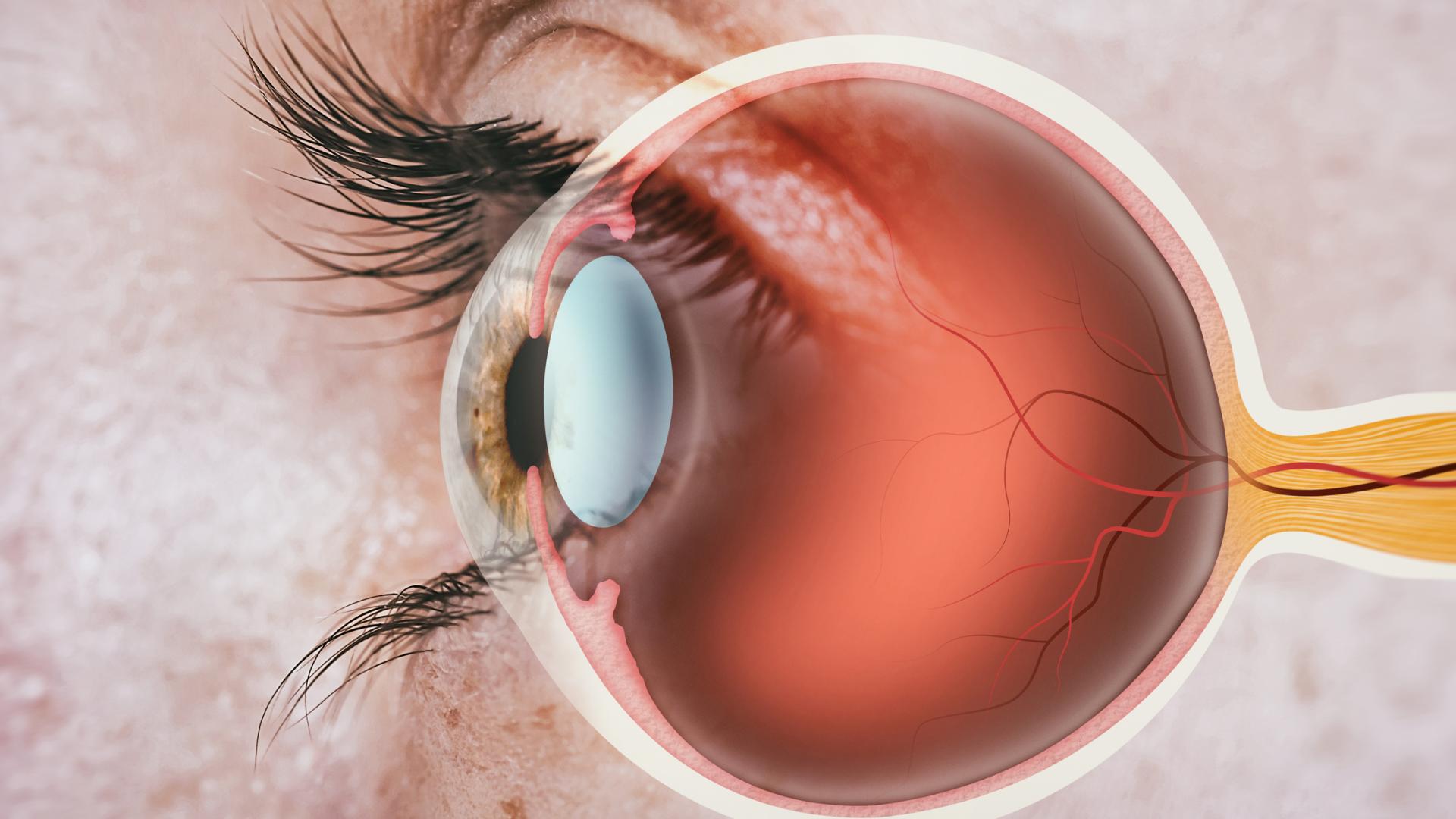
DR. LAUREN HOCK: Yeah, this test is so important, and it’s really an artform to help us know how the drain in your eye is working. Gonioscopy is performed with a handheld lens, and some doctors, including me, refer to it as a contact lens. This is because we numb up your eye and then barely touch the lens to the surface of your eye to take a look at the angle between your cornea, which is the clear part in the front part of your eye, and the iris, which is the part of the eye that gives it its color. The procedure is simple, quick, and it doesn’t hurt. And it tells us whether the fluid that’s created in your eye has room to exit the eye by passing through the drain, which is another word for the trabecular meshwork. If, when you look with that contact lens, your doctor can see your trabecular meshwork, you have open-angle glaucoma, which is treated one way. And then if, when your doctor looks, they can’t see your drain or your trabecular meshwork, this is called narrow or closed-angle glaucoma. And the treatments for these diseases can be quite different. Some people who have been referred for narrow-angled or closed-angled glaucoma can expect to have this test repeated more frequently if their doctor is determining whether they might need a laser or something like cataract surgery to help create some more space in their eye.
MS. KACI BAEZ: Thank you. And so, how frequently should somebody diagnosed with glaucoma be tested with some of these exams in comparison to the frequency guidelines for a screening exam? And how often should eye pressure be checked?
DR. LAUREN HOCK: It really depends, is the answer. And it’s definitely going to be more frequent overall if you have glaucoma—particularly if you have moderate or severe glaucoma than if you’re someone who’s just been determined to be high risk for glaucoma. Some tests—like pressure measurement; testing your visual acuity, which is how well you read the eye chart; and then looking at you with the microscope or the slit lamp—should be performed at every visit. Most glaucoma specialists will check a visual field test and an OCT at least once per year. Although, if there’s concern that there’s changes to your vision or changes to your glaucoma, these may be done more frequently. And then, there’s some evidence that there’s a small percentage of people whose glaucoma will get worse more rapidly, and we can help detect who those people are earlier by performing more visual field tests in the first 2 years. Again, that’s a small portion of people, but some glaucoma doctors will do the glaucoma field test more frequently when you’re first diagnosed, trying to make sure that you’re in the larger category of people whose glaucoma will change very slowly over time.
MS. KACI BAEZ: Thank you. Just thinking about these different tests, how would somebody prepare for an eye exam, and how long would they typically take?
DR. LAUREN HOCK: Yeah, that’s a great question. So, most tests for glaucoma should take no more than a few minutes, and there’s really no real special preparation for these tests, but I don’t want anyone to get too psyched out about a visit to their glaucoma specialist. There’s different ways of doing the visual field test and different machines, but usually, they’ll only take a few minutes per eye, and then the photo tests are quite quick. Once you look in the right spot, it shouldn’t take more than a few minutes. With the visual field test, if it’s taking a really long time or your doctor notices that you’re having trouble concentrating for the whole test, there’s different strategies or approaches for doing these tests, and they can make some adjustments to better set you up for success. It’s worth knowing that if your doctor plans to dilate your eyes, you should expect a little bit longer of a visit. Most people will wait at least 10 to 20 minutes for your pupils to dilate so they can get a good look at the health of your retina. And then just in general, if you’re someone who’s lost a significant amount of vision from glaucoma or other eye problems, some tests may take you longer to perform, and that’s okay. We really just want to make sure that we’re getting the best information about your eye health that we can so we can tailor your treatment.
MS. KACI BAEZ: Great. So, are there any possible risks or side effects from a glaucoma eye exam that our listeners should know about?
DR. LAUREN HOCK: That’s a great question, too. So, some of my patients experience dryness or irritation from the numbing drops that we use. Usually that goes away relatively quickly, but people can always use lubricating drops if they’re having issues after the glaucoma exam. There’s really very few risks of these exams unless you’re someone who’s been told you have narrow angles or angle-closure glaucoma. In certain situations, your doctor may choose to not dilate your eyes or may caution you against having a dilated eye exam while you’re being evaluated. But there’s very few side effects, and there’s very little preparation other than trying to get some rest the night before.
MS. KACI BAEZ: That’s really helpful. So, always check with your doctor for specific questions about tests, but can you drive after a glaucoma eye test?
DR. LAUREN HOCK: Most of my patients who do drive, drive themselves to their appointments. Especially if you’re not having a dilated eye exam, aside from a little bit of irritation, you should expect to feel pretty normal after your eye exam. I will say that some people with glaucoma have lost some of their vision and may just have difficulty driving in general, and that should be discussed specifically with their eye doctor. And then, if your eyes are dilated, some patients feel comfortable driving and some don’t, but if you know you’re in the category where you feel kind of funny after your eyes are dilated, it’s not a bad idea to bring a driver.
MS. KACI BAEZ: Thank you. “Can cataracts interfere with the glaucoma eye exam?” is another question our listeners have.
DR. LAUREN HOCK: Yeah, that’s a great question. So, most glaucoma specialists take care of cataracts, too, and they can definitely affect your eye exam. In general, cataracts are usually fully progressive and can blur your vision, so once they get to be more significant, they can affect your ability to see more clearly, and that can make you perform a little bit worse on the visual field test. If your cataracts have grown a lot, sometimes it can affect our ability to see from the outside of the eye into your eye and assess your glaucoma that way, at which point many people would recommend cataract surgery. And then, I think the final important way that’s less common but certainly seen in the glaucoma clinic is sometimes people’s cataracts can contribute to a crowding of that angle we talked about in the eye, and so some people’s doctors will recommend cataract surgery a little bit earlier than their friends and family may have had it done if that’s a concern specific to them.
MS. KACI BAEZ: Okay, great. And so, if someone has gotten these eye exams and they want to see their results, are they able to see their results? Understanding that they’re best reviewed with their doctor, but if someone really wanted to, you know, interpret their results and view them, is that possible?
DR. LAUREN HOCK: Yeah, it should be possible. Things like your visual acuity and your eye pressure and the actual exam that your doctor sees will be written down in their chart notes, and that can be very easily made available as a printout or electronically. And then, the nice thing about doing these tests with the visual field and the OCT are that these are easily followed either electronically or by printouts over time. Many of my patients who’ve maybe been followed in another state or elsewhere in the community and will come to my office, Wills Eye Hospital, will bring printouts of their old tests. And it’s really helpful to look at these together to see, “Okay, what was your glaucoma like 10 years ago? Has everything been super stable? You have no changes to your vision; that’s great. Maybe we can relax a little bit,” or “Wow, it seems like things have really changed over the last 5 years. Maybe we need to rethink how we’re treating your glaucoma.” The results, in terms of the numbers, sometimes can change from machine to machine, and so it’s helpful to review those specific numbers with your doctor, but the actual printouts are usually able to be made available pretty easily. And I guess the final portion of that was, as far as an electronic portal, that’s probably going to depend on each doctor’s individual office as to what is visible on a portal, so you should ask your specific eye doctor’s practice how they handle that.
MS. KACI BAEZ: Okay, great. Knowledge is power.
DR. LAUREN HOCK: Yes.
MS. KACI BAEZ: And so, would you say there’s one test that is more useful than the others in terms of these glaucoma eye exams, these six tests we’re talking about today?
DR. LAUREN HOCK: Sure. I think if I had to choose one test that I could do each time, I guess it would be checking your vision and checking your pressure, since that’s the thing that can most determine adjustments to your treatment. Each of the other tests provide slightly different information that helps us know how you’re doing and how the glaucoma is impacting your life. So, in terms of the visual field test, that’s really a great test for telling us: How is the glaucoma affecting your ability to function out in the world? Do we think it’s affecting your driving? If you’re missing some of your vision kind of below the center of your vision, are you missing things on the ground? Are you at risk for falling? Do we need to talk about figuring out ways to keep you safer at home? But really, each of the tests kind of provides their own specific thing, but vision and pressure are the ones that we’re going to check every visit.
MS. KACI BAEZ: Okay, thank you. Can these tests be normal and someone still have glaucoma?
DR. LAUREN HOCK: That is an excellent question. Some of these tests results can fluctuate a little bit from visit to visit. Some tests, such as the visual field test, we actually expect to be normal early in glaucoma. People with mild glaucoma often will have no symptoms of vision change and only have structural changes on some of those object nerve imaging tests that we discussed. So certainly, some of the tests can be normal, but you might still benefit from treatment to reduce the risk of ever losing vision from glaucoma. And then, some people who have a family history or have something called narrow angles like we talked about, you might have borderline findings that are mostly normal, but your doctor might be worried will change in the near future and might continue to monitor you closely to detect if anything becomes abnormal so they could catch it early and get you treated.
MS. KACI BAEZ: Great. Thank you. We talked about the home eye pressure device, and there’s been an uptick in at-home testing interest, and another question we have is: Is there a blood test for glaucoma?
DR. LAUREN HOCK: Yeah, that’s a great question. To my knowledge, there’s no single blood test available to diagnose glaucoma. There are definitely researchers who look at biomarkers for glaucoma in the blood, but these are not commonly used in day-to-day clinical practice. The exception for this would be if you’re someone who was diagnosed with glaucoma as a child or as a young person in your teens, 20s, or even 30s, it might be helpful to meet with a genetic counselor or talk to your doctor about genetic testing to understand you and your family’s risk for glaucoma. There are specific genes that can run in families and cause people to develop glaucoma at an earlier age, so it can be really helpful if you’ve had a diagnosis of glaucoma at a young age to get some blood work done so you know whether your siblings or other family members might be at risk. In general, doing a test that you can just order straight from yourself, like 23andMe, there’s not like a specific blood test that can tell you definitely you will or will not develop glaucoma. And because multiple genes can be at play, routine genetic testing for people who develop run-of-the-mill glaucoma at an earlier age is not recommended.
MS. KACI BAEZ: Thank you, and it’s so interesting. I know I personally have a family history of glaucoma, and I did get one of those at-home genetic tests, and it did confirm that glaucoma—
DR. LAUREN HOCK: Oh, really?
MS. KACI BAEZ: Yeah, I’m at a higher risk. But we’d obviously never use that in place of going to the doctor, right, and getting an eye exam, because half of the 80 million people across the world who have glaucoma don’t know they have it.
DR. LAUREN HOCK: Right.
MS. KACI BAEZ: It is quite interesting, with the uptick of new tools available for people on their personalized medicine journey.
DR. LAUREN HOCK: Yeah, I think this is a really exciting area of research that’s getting closer and closer to being able to be brought into the clinic, but certainly having a sense of, “Oh, you have this gene and this gene, and maybe you’re at higher risk for getting glaucoma at an earlier age, so we should follow you more carefully.” But in terms of diagnosing glaucoma, we really need to look at how you’re doing and what your eyes actually look like to know for sure how you’re doing.
MS. KACI BAEZ: Okay. It is eye exam awareness time in January. Glaucoma awareness.
DR. LAUREN HOCK: Yes. Absolutely.
MS. KACI BAEZ: Everybody, go visit your doctor.
MS. KACI BAEZ: One of our final questions is: Is there any research that’s being done to help in terms of diagnostic tools or glaucoma exams or anything in development or on the horizon that we should know about?
DR. LAUREN HOCK: Yes, absolutely. And I think the question about, “How can we capture some of the changes people are experiencing with their vision and their glaucoma at home?” is one of the most exciting areas in development that I think will be more home based in the somewhat near future. We talked about the iCare HOME, but there’s also ways to just check your visual acuity at home and with lower technology demands like with home applications with that standardized eye chart that’s used instead of community care all over the world. For checking pressures at home, we mentioned the iCare HOME. One of the ways I use this with my patients is our office actually will rent or loan these devices to patients. In my clinic, while I might be worried that something’s changing, “Their pressure is always normal in clinic, but it seems like their glaucoma is getting worse. What’s going on?” And sometimes, they’ll come back with these readings where the pressure is maybe 10 to 15 points higher than I’ve ever measured it in clinic, and so we know, “Wow,” you know, “We’re under-treating this.” There’s also some wearable devices in development. There’s a contact lens called the Triggerfish that I haven’t used personally but detects pressure fluctuations by placing a contact lens on the eye. In Europe, I believe, there’s an implantable device that would be surgically placed. It’s a microsensor that detects changes in pressure, but this is not quite available in the United States yet.
And then, a lot of different academic groups and groups in technology are working on virtual reality headsets for doing the visual field testing. So, there’s already tablet-based visual field tests available—which are great, but sometimes the lighting conditions can be variable—but when you place these headsets on, we can better control the amount of light your eye is perceiving and some of the reliability of that test. There’s also work done with portable camera imaging, either a smartphone or even a home-based OCT, which is that quantitative test that we talked about. I haven’t personally used a home OCT with any of my patients yet, but I think it’s a really exciting area of investigation. It’s Glaucoma Awareness Month. We want to connect to all those people who have glaucoma and don’t know about that, so all of these tests that we can do outside of the clinic in the community setting, whether at home or perhaps at a primary care office, will be so helpful in detecting people who are at risk for losing their sight and should be connected with a glaucoma specialist. And then, just to kind of tie that off, artificial intelligence is an exciting thing that’s affecting all parts of society, including medicine, and there are several research groups that are looking at how to detect glaucoma earlier and catch progression using algorithms with artificial intelligence. There’s some commercially available screening devices for retinal diseases, and we’re hoping that artificial intelligence can enhance our ability to detect glaucoma with those similar devices.
MS. KACI BAEZ: Wow, well the power of research is truly incredible. It’s so exciting to hear about all these developments on the horizon. Thank you, Dr. Hock, for sharing that with us.
DR. LAUREN HOCK: Yeah, of course. Yeah, we have a study at Wills Eye Hospital where we’re trying to catch some of these changes at home, and there are certainly many excellent groups here in the U.S. that are working on this. Not everyone uses the same devices, but we all share a common goal of trying to catch changes in glaucoma with people being in the comfort of their own home, not needing to drive to the eye exam every time, take time off work, or get a family member to drive them. And then, maybe we can give more tailored treatment with that additional information, too.
MS. KACI BAEZ: So many exciting developments that have just come out over recent years due to research or are in development. This is just so wonderful and hopeful. Those were all the questions we had today, Dr. Hock, unless there’s any last information you’d like to share with our listeners.
DR. LAUREN HOCK: I don’t think so. I just would encourage any of our listeners who have a diagnosis of glaucoma to please encourage your first-degree relatives—your parents, siblings, and children—to get a comprehensive eye exam. We know that the earlier glaucoma is caught, the more we can do to reduce the risk of people losing their sight, and we want everyone to lead happy, healthy lives with the best vision that they can have.
MS. KACI BAEZ: Wonderful. Well, thank you, and so, I hope—our listeners—that you found our time together today helpful. Our next Glaucoma Chat will be on Cataracts and Glaucoma: What You Should Know in 2024 and will be live on Wednesday, February 14—Valentine’s Day lunch treat. Thank you again for joining us, and this concludes today’s BrightFocus Glaucoma Chat.