


Diagnosis & Testing
Early detection and treatment of glaucoma can slow the progression of the disease. Vision loss due to glaucoma is irreversible and, if left untreated, can lead to blindness. To avoid permanent vision loss, it is important to have your eyes examined for glaucoma by an eye doctor. Regular eye exams are especially important if you carry known risk factors, such as a family history of the disease.
Support Vision-Saving Research
More than 4 million Americans are living with glaucoma. Your gift can help fund research that could protect their sight and provide vital information to the public.
What to Expect
If you or a loved one has just been diagnosed with glaucoma, it can be a stressful time for you and your family. Here are a few tips and things to expect after receiving a diagnosis:
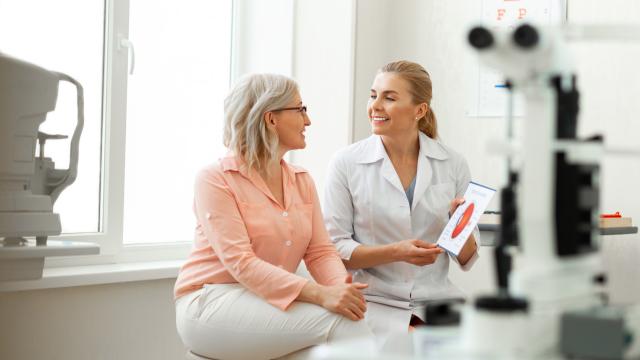
Find an Eye Doctor You Trust
You will have an increase in doctor appointments, so finding an eye doctor you feel comfortable with is important. Coordinate your treatment plan with your support team, and remember to prepare for your appointments to make the most of the time spent with your doctor.
Prepare for Potentially Increased Medical Costs
Like with any chronic disease, medical costs can be high. Learn about federal, state, and local government benefits programs to help with costs and find other resources.
Additional Testing
Further testing, such as optic nerve imaging, may be needed to confirm your diagnosis or determine the stage of the disease. Continue reading for more information about screening and testing.

Types of Testing
If you’re at high risk for glaucoma, you should have a dilated eye examination at least every one to two years. To help diagnose glaucoma, an ophthalmologist or optometrist will perform a comprehensive eye exam that could include a range of tests.
Eye Pressure Check
Increased eye pressure is the most important risk factor for glaucoma. The eye pressure test, called tonometry, is the most common method of checking for increased eye pressure. There are several methods of measuring eye pressure. The most common method is known as applanation, in which a tiny instrument contacts the eye’s surface after it is numbed with an eye drop.
Visual Field Test
This test measures the entire area the forward-looking eye sees to document straight-ahead (central) and side (peripheral) vision. It measures the dimmest light seen at each spot tested. Each time patients perceive a flash of light, they respond by pressing a button.
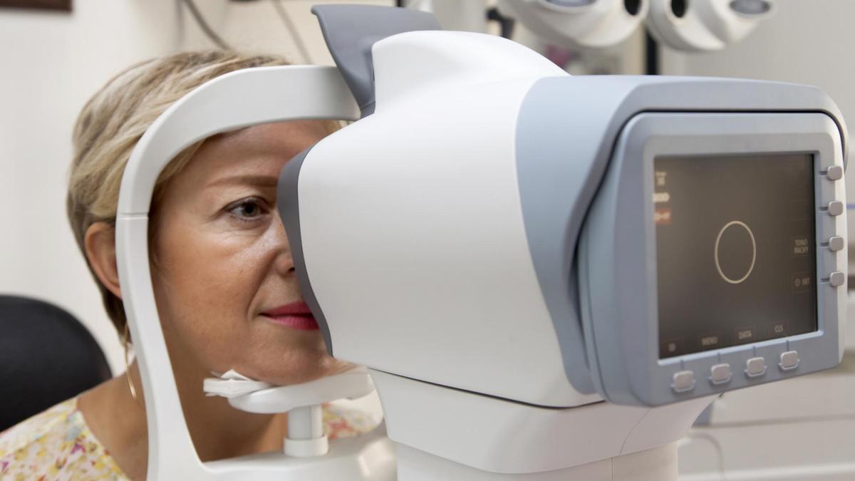
Glaucoma Imaging Tests
After dilating your pupils using eye drops, the doctor will photograph your optic nerve to map and document the health of your optic nerve.
Dilated Eye Exam
This test uses special eye drops that temporarily enlarge the pupil so that the doctor can get a magnified, 3D view of your optic nerve and check for diseases like glaucoma.
Visual Acuity Test
This test measures sight at various distances. While seated 20 feet from an eye chart, the patient reads standardized visual charts with each eye, with and without corrective lenses.
Cornea Thickness Test (Pachymetry)
The eye doctor uses an ultrasonic wave instrument to help determine the thickness of the cornea and better evaluate eye pressure.
Ophthalmoscopy
The doctor examines the eye’s interior by looking through the pupil with a special instrument. This test can help detect damage to the optic nerve caused by glaucoma.
Angle Test (Gonioscopy)
This test allows the eye doctor to see the “angle” where the cornea meets the iris. This is also where the trabecular meshwork (the eye’s drainage system) is located. This test can help diagnose closed-angle glaucoma.
Optic Nerve Imaging
Imaging helps document optic nerve changes over time. Nerve imaging techniques include:
- Stereo optic nerve photographs
- Scanning laser polarimetry (GDx)
- Confocal scanning laser ophthalmoscopy (Heidelberg Retinal Tomograph or HRT)
- Optical coherence tomography (OCT)
All four techniques are painless and noninvasive. Your doctor will determine which method(s) to use, depending on your glaucoma condition.
Read more about eye exams and testing for glaucoma using our helpful resource.
What is a Glaucoma Suspect?
A glaucoma suspect is an individual who demonstrates one or more factors that put them at higher risk of a glaucoma diagnosis, but do not yet have glaucoma damage. Sometimes this is referred to as pre-glaucoma or borderline glaucoma. Characteristics of a glaucoma suspect include:
- High intraocular pressure (IOP) or ocular hypertension
- Unusual or defective visual fields
- Other optic nerve features suggestive of glaucoma
If you are a glaucoma suspect, the most important treatment is good follow-up care. Sometimes eye doctors are on the fence about whether to start treatment, and it is only through repeat follow-up visits that they get a sense of whether or not someone has glaucoma. Thus it is very important to maintain follow-up care. Typically for a low-risk glaucoma suspect, this may require visits every 6 to 12 months.
There is Hope
National Glaucoma Research, a BrightFocus Foundation program, is on a mission to stop vision loss in its tracks. For the last 50 years, we’ve funded the boldest research and “what-if” ideas to get us closer to cures, resulting in the novel treatments and diagnostic tools in use today.
Confused by medical lingo?
Were you or a loved one recently diagnosed with glaucoma but feel lost when trying to decipher the diagnosis given by your physician? Use our disease glossary to learn more about the terms you’ve been hearing.

Search for a Glaucoma Clinical Trial
Clinical trials are crucial to advancing the most effective medical approaches. Today’s studies will lead to new standards of care in the future.

Resources
Recent Resources & Information
Downloadable Resource
The Top Five Questions to Ask Your Eye Doctor
Preparing ahead of time can help you best manage your vision health. Here are some questions you can take along when you visit the eye doctor.

Glaucoma Chats
Gathering Support For Your Glaucoma Diagnosis
Join Dr. Lawrence S. Geyman as he outlines effective ways to find support for your glaucoma diagnosis.

Glaucoma Chats
Glaucoma and Macular Degeneration: Can You Be Diagnosed With Both?
Learn about the similarities and differences between these two vision diseases, whether someone can develop both conditions at the same time, and how to reduce your risk.
Podcast
Childhood Glaucoma
Learn about the different types of childhood glaucoma, including primary congenital, infantile, and juvenile glaucoma.
Glaucoma Chats
Exploring Artificial Intelligence and Glaucoma: Challenges, Opportunities, and Hope
Discover the innovative ways scientists and healthcare professionals are leveraging artificial intelligence to detect and diagnose glaucoma.
Podcast
Glaucoma Imaging: Trends in Detection and Diagnosis
An expert outlined the latest advancements in optic nerve imaging that have enabled earlier glaucoma detection.