Resources > Expert Information
Published:
National Glaucoma Research
Pupil dilation is performed to purposefully increase the size of the pupils during an eye exam so that the eye doctor can fully examine the health of the optic nerve and retina. The exam is critical to preventing and treating eye conditions that could potentially lead to vision loss.
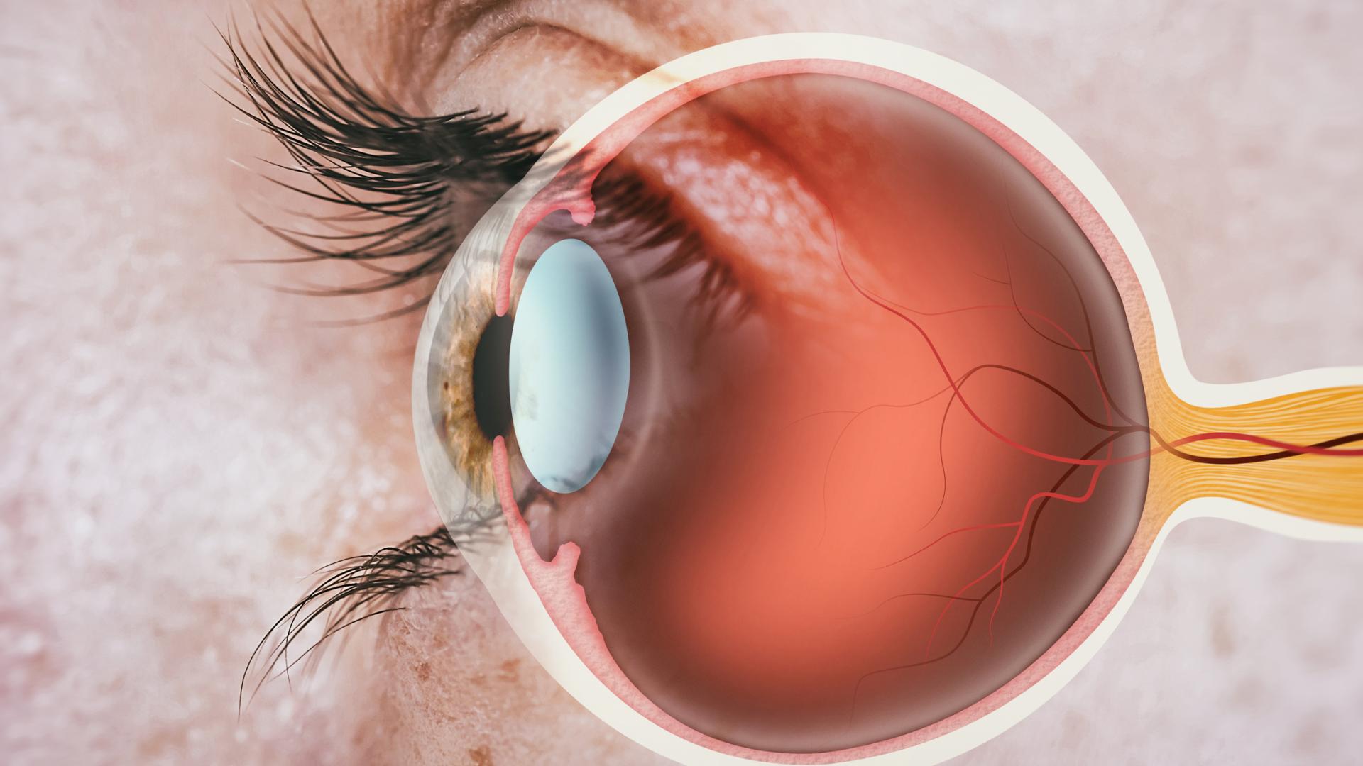
Pupil dilation occurs when the opening in the center of your iris grows bigger to let in more light. Under normal circumstances, pupils can dilate to let in more light or in response to a variety of stimuli. During an eye exam, a doctor will administer eye drops to increase the size of a patient’s pupils. The eye is a beautiful organ, and it is the only place in the human body where a doctor can see a part of the central nervous system, the optic nerve. Seeing the optic nerve is a crucial part of a comprehensive eye examination.
Both dilated and undilated eye exams provide important information to an eye doctor. Let’s explore the undilated exam first.
One of the first parts of a comprehensive eye exam is a test of your vision, and perhaps a measurement to determine an eyeglass prescription, both of which require that your eyes remain undilated.
In addition, eye doctors will examine your pupils’ responses to light prior to dilation. This can be important for determining whether the visual pathways for each eye are working properly.
Next, the eye doctor may use a special microscope called a slit lamp to examine the front of your eyes. This includes looking at the eyelids, the cornea or clear “window” front part of your eye, the iris or round colored part of your eye, and the lens, which is a major part of the eye giving it the ability to focus.
There is also an examination, called gonioscopy, which allows the doctor to examine your eye’s drainage angle with a special mirrored lens. The “angle” that is being referred to is the angle between the iris and the cornea, which are both described above. When the angle is open, your ophthalmologist can see most, if not all, of your eye’s drainage system. When the angle is narrow, only portions of the drainage angle are visible, and in an acute angle-closure event, none of it is visible.
Part of a glaucoma examination is formal visual field testing, where your peripheral, or side vision, is tested. Ideally, your eyes are not dilated during this test.
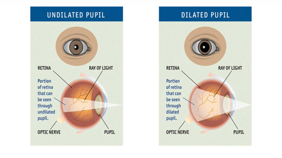
The view to the back of the eye is limited when the pupil is not dilated. When your pupil is small, an eye doctor can see your optic nerve and macula but the view is limited. In order to see the entire retina, the pupil must be dilated. This is achieved by placing dilated drops in the eyes.
Once the drops are administered, it typically takes 15-30 minutes to achieve fully dilated pupils, depending on the person’s response to the medication.
When eyes are dilated during an eye exam, it typically takes 4-6 hours for pupils to return to normal. Some individuals may experience eye dilation that lasts longer.
Once your eyes are dilated, there is an increase in light sensitivity because the pupil is large and more light is coming through, so bring your sunglasses, or your ophthalmologist may provide some disposable shades for your use. You may also experience blurry vision, particularly if you are trying to read. Some patients feel a “tightening” or different sensation in their eyelids. If it is your first time having your eyes dilated or you know your vision is too impaired for driving after dilation, bring a friend or companion to drive you home from your examination. While in the past there were some eye drops that could reverse the dilation, these are no longer available, so you will have to wait the 4-6 hours before the drops completely wear off.
The optic nerve can be seen through an undilated pupil, but for optimum viewing a dilated pupil is required. This is important for the diagnosis of glaucoma, as well as other diseases of the optic nerve. Learn about what to expect during a glaucoma eye exam.
Two very common retinal diseases, diabetic retinopathy and age-related macular degeneration (AMD), are diagnosed and monitored by examining the retina through a dilated pupil. Learn about what to expect during a macular degeneration eye exam.
In addition to macular degeneration and glaucoma, there are many other conditions that require pupil dilation, such as detection of a retinal tear or detachment, or an ocular tumor, just to name a few. Finally, to fully see a cataract, or clouding of the lens, a dilated eye is helpful.
As part of a comprehensive eye examination, pupil dilation is very important to reveal the status of your optic nerve and retina and is critical to preventing and treating eye conditions that could potentially lead to vision loss.
BrightFocus Foundation is a premier global nonprofit funder of research to defeat Alzheimer’s, macular degeneration, and glaucoma. Since its inception more than 50 years ago, BrightFocus and its flagship research programs—Alzheimer’s Disease Research, Macular Degeneration Research, and National Glaucoma Research—has awarded more than $330 million in research grants to scientists around the world, catalyzing thousands of scientific breakthroughs, life-enhancing treatments, and diagnostic tools. We also share the latest research findings, expert information, and resources to empower the millions impacted by these devastating diseases. Learn more at brightfocus.org.
Disclaimer: The information provided here is a public service of BrightFocus Foundation and is not intended to constitute medical advice. Please consult your physician for personalized medical, dietary, and/or exercise advice. Any medications or supplements should only be taken under medical supervision. BrightFocus Foundation does not endorse any medical products or therapies.

Encontrará recursos e información que ofrecen productos, servicios y otro tipo de apoyo para personas con glaucoma y sus familias.
In clinical trials, stem cell therapy for glaucoma shows promise for rebuilding the eye’s drainage system and protecting the optic nerve.

Meet Richard, a loyal donor to BrightFocus' National Glaucoma Research and Macular Degeneration Research programs.

Resources and information that provide products, services, and other support for people with glaucoma and their families.
Support Groundbreaking Glaucoma Research
Your support helps fund critical research that could prevent vision loss, provide valuable information to the public, and cure this sight-stealing disease.
Donate Today