Resources > Expert Information
Macular Degeneration Research
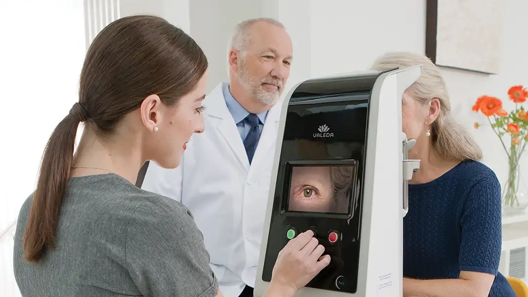
The goals of a typical visit to the ophthalmologist for macular degeneration are to confirm or refute the diagnosis, assess future risk, and provide any required treatments. The exam includes several components, including the history, examination, and computerized imaging. The entire process including exam with pupil dilation, imaging and treatment can take several hours.
Your eye doctor will ask questions about your vision by taking a medical history and conducting a comprehensive eye evaluation.
During your eye exam, your ophthalmologist will ask questions, such as:
Remember to bring your medicines or a list of them with doses to your first appointment. Also, bring the eyeglasses you use the most often and like the best.
Usually a technician will check your vision on an eye chart with and without glasses, and may test whether your glasses need adjustment by testing you with the phoropter. This is the device containing lots of lenses. When you look through the phoropter, the technician will change the lenses and ask you “which looks clearer, number 1 or 2?” If they look about the same, just say so. That’s a helpful answer too.
The technician may also check your eye pressure to screen for glaucoma. During this test, yellow eye drops will be put in your eyes and you will be asked to keep your eyes open while a meter (called a tonometer) with a blue light measures your pressure. You won’t feel anything during this test; however, it is important not to rub your eyes for 10 minutes after the eye drops are used, as it is possible that you might inadvertently scratch the front of your eyes (your corneas) if you rub them while your eyes are numb. You can close your eyes and dab gently with a tissue, however.
Finally, the technician will put eye drops in your eyes to dilate your pupils so that the doctor will have a good view of your retina. If you have brown eyes, your pupils usually remain dilated for about four hours. For blue eyes, the pupils may remain dilated until the following day. Remember to bring sunglasses and, ideally, someone to drive you home after your appointment, as you will be sensitive to light.
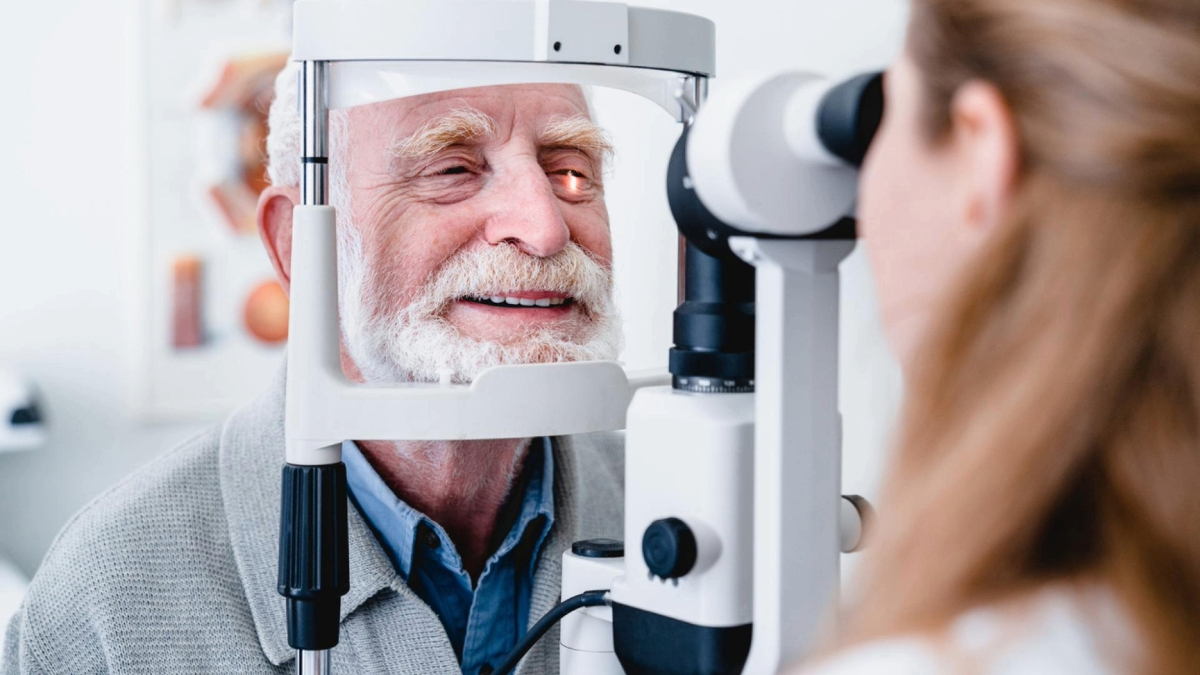
About 30 minutes after the eye drops are put in, your pupils will be fully dilated, and the eye doctor will examine you. The doctor may move his/her finger in front of your eyes, and you will be asked to follow the finger so that your eye movements can be observed. The doctor may also put their hand in different positions and ask you how many fingers you see to test the different parts of your visual field. Then, they will ask you to put your chin on a chinrest and your forehead against a strap while they examine your eyes with a slit lamp, which is a microscope with a moving light. At times, the light may seem bright, but try to look straight ahead and keep your eyes open as much as possible. It’s fine to blink briefly. This part of the exam usually only lasts a minute or so.
The doctor may then put a light on their head and ask you to look in several different directions while they observe the edges of your retina with an indirect ophthalmoscope. Again, the light may be bright, but the exam won’t last long. Sometimes, when asked to “look left,” a patient will ask, “with which eye?” Remember that both eyes move together, so they will both be moving in the same direction.
For macular degeneration exams, imaging often includes retinal imaging and optical coherence tomography (OCT). The retinal photographs consist of a few bright flashes of light in each eye, and only take a few minutes. In OCT, you’ll see a small flashing light in the middle of your field of view to help show you where to look, but there are no bright lights. The exam takes about one minute per eye. The OCT provides a beautiful cross-section view of the retina, so the doctor can assess its structure and see whether any fluid has leaked from abnormal vessels and accumulated in the retina.
If the doctor thinks there may be wet macular degeneration, he/she will order a fluorescein angiogram or indocyanine green angiography. These tests involve injection of a dye into an arm vein followed by timed photographs of each eye at intervals for about 15 minutes following the injection. This enables the doctor to see if any dye is leaking out of the abnormal blood vessels in the retina that might require treatment to protect your vision. These tests are generally very safe, but occasionally some of the dye leaks out of the vein and into the area under the skin near the injection site. This can cause arm pain that is managed with an ice pack. People, who tend to be allergic to many things, including shellfish, may have an allergic reaction to the dye. If you tell the doctor about your allergies, he/she may give you a Benadryl prior to the dye injection to decrease the chance of an allergic reaction. The fluorescein angiogram will cause your urine to turn a very bright yellow for about one day, as the fluorescein is eliminated from your body.
In this era of digital, computerized tests, the eye doctor can often give you your test results within a few minutes, and then let you know whether you need a treatment, such as injection of brolucizumab (Beovu®), aflibercept (Eylea®), ranibizumab (Lucentis®), or (bevacizumab (Avastin®) for wet macular degeneration. If you have dry macular degeneration, the treatment recommendations may include vitamins, dietary modification, and follow-up visits to monitor your retinal health.
It is important to remember that with modern exam techniques and treatments, many people with macular degeneration keep good vision for their entire lives.
BrightFocus Foundation is a premier global nonprofit funder of research to defeat Alzheimer’s, macular degeneration, and glaucoma. Since its inception more than 50 years ago, BrightFocus and its flagship research programs—Alzheimer’s Disease Research, Macular Degeneration Research, and National Glaucoma Research—has awarded more than $300 million in research grants to scientists around the world, catalyzing thousands of scientific breakthroughs, life-enhancing treatments, and diagnostic tools. We also share the latest research findings, expert information, and resources to empower the millions impacted by these devastating diseases. Learn more at brightfocus.org.
Disclaimer: The information provided here is a public service of BrightFocus Foundation and is not intended to constitute medical advice. Please consult your physician for personalized medical, dietary, and/or exercise advice. Any medications or supplements should only be taken under medical supervision. BrightFocus Foundation does not endorse any medical products or therapies.

Gene therapy treatments may one day free people with age-related macular degeneration (AMD) from the need for frequent eye injections.

Dave and Leanna Palmer share their commitment to supporting Macular Degeneration Research.
Artificial vision systems utilizing these technologies show promise for individuals with profound vision loss in early trials.

Promising new treatments in late-stage clinical trials could transform how we treat age-related macular degeneration.
Help Fight Macular Degeneration and Save Sight
Your donation helps fund critical research to bring us closer to a cure for this sight-stealing disease and provide vital information to the public.
Donate Today