Focusing on the Future: Key Takeaways from the 2025 Glaucoma Fast Track
BrightFocus Foundation’s 2025 Glaucoma Fast Track brought together early-career and established scientists to provide a thorough overview of the biology, diagnosis, and treatment of glaucoma.
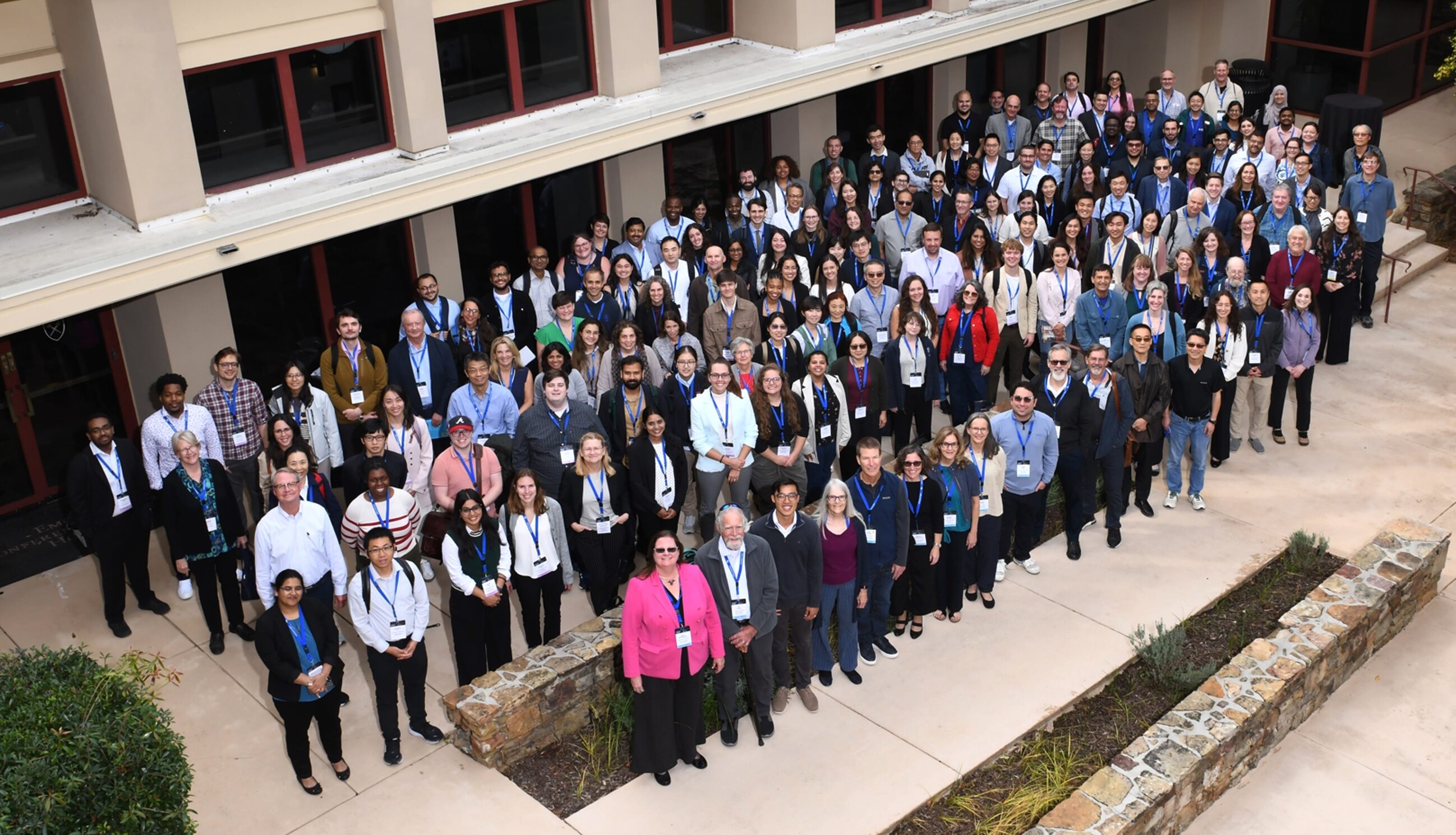
2025 Glaucoma Fast Track participants
On October 8, 2025, BrightFocus Foundation presented the 4th Glaucoma Fast Track in Atlanta, GA, as the official pre-symposium workshop to the International Society for Eye Research (ISER)/BrightFocus Glaucoma Symposium: Concepts and Breakthroughs in Glaucoma. The Fast Track was attended by more than 127 clinicians and researchers from 7 countries on 4 continents, reflecting the global scope of the glaucoma research community and the broad interest in this educational workshop. This year’s workshop was co-chaired by Diane Bovenkamp, PhD; Abbott F. Clark, PhD; Colleen McDowell, PhD; and Rebecca M. Sappington, PhD.
This year, BrightFocus offered a special pre-workshop session on scientific communication and collaboration exclusively for travel fellows the day before the Fast Track. Bri McWhorter, founder and CEO of Activate to Captivate, led an interactive workshop focused on helping scientists distill complex concepts into clear, engaging narratives to share with funders, other scientists, and individuals affected by disease. Fellows then had the opportunity to apply these skills by presenting their innovative research through oral and poster presentations at the ISER-BrightFocus Glaucoma Symposium, which took place immediately after Fast Track.
BrightFocus Foundation’s Fast Track workshops, offered across Alzheimer’s disease, age-related macular degeneration, and glaucoma, bring together established leaders in the field and early-career scientists to promote mentorship, education, and the exchange of ideas. A strong emphasis is placed on creating an inclusive and welcoming environment where young investigators feel empowered to ask questions and strike up conversations with their senior colleagues.
In her opening remarks, Diane Bovenkamp, PhD, vice president of scientific affairs at BrightFocus, emphasized the importance of learning from each other and taking advantage of networking opportunities provided by the Fast Track:
“We started Fast Tracks as a ‘boot camp’ for early-career investigators. I encourage everybody in the room, especially our 56 travel fellows, to just walk up to anyone. Even if they’re probably going to win the Nobel Prize next year, don’t be afraid to walk up to them and ask them questions. They would love to talk with you about your work.”
To date, the program has trained 181 early-career investigators from 16 countries. Fifty-six young investigators attended the workshop thanks to a BrightFocus travel award.
56 early-career investigators received a BrightFocus travel award to help defray the cost of attending this year’s Glaucoma Fast Track.
Presentations thematically moved from glaucoma fundamentals to emerging technologies and future therapies. The opening presentation was given by Dr. Yvonne Ou, ophthalmologist and researcher at the University of California, San Francisco, whose research has been supported by a BrightFocus Foundation National Glaucoma Research grant. She provided a broad overview of glaucoma, its diagnosis and treatment, and what gaps remain in patient care.
In subsequent sessions, speakers gave overviews on the following topics:
Fundamentals: the strengths and limitations of experimental glaucoma models, emphasizing that studies carefully designed around known limitations provide valuable mechanistic insights.
New Models and Modeling Outcomes: highlighting innovative eye imaging techniques and powerful new technologies using stem cells derived from people with glaucoma. One presentation showcased modern twists on “eyes in a petri dish,” first pioneered in 1880.
Artificial Intelligence (AI) and Imaging: approaches for leveraging AI methods for detecting glaucoma and predicting the disease course.
Genetics and Future Therapies: the identification of risk genes for glaucoma and considerations for potential future gene therapies.
Financing Scientific Pursuits/Goals: a panel discussion that provided advice on career paths and funding strategies, encouraging resilience in the face of rejection.
Five Key Takeaways
Don’t Forget the Human Side
Eye exams can be tiring and uncomfortable. Despite this, regular, frequent eye exams are important, especially for people diagnosed with elevated intraocular pressure (IOP) and glaucoma. If IOP is monitored at regular intervals, it can often be kept in check with medication or surgery, slowing progression and preserving vision. At the same time, measuring IOP alone is not enough, it is also crucial to determine if there are associated changes in vision.
Dr. Yvonne Ou underscored this point in her opening presentation when she shared the story of a patient with elevated IOP who had to switch providers because of insurance changes. She eventually saw him again after eight years, but unfortunately his vision had deteriorated significantly. She explained that his progressive vision loss could have been detected sooner, and possibly treated more effectively, if he had been examined more frequently: “If you do one test per year, it’s going to take you four years to detect that progression. If you do two tests per year, you decrease that time to detection to three years.”
Ultimately, getting regular eye exams is vital for the early detection and effective treatment of glaucoma, but life is never perfect and things get in the way, both on a human level – fear of uncomfortable medical procedures, anxiety about results – as well as factors beyond a patient’s control such as insurance issues. Optimal patient care runs up against these real-life curveballs, and it is important for caregivers to be aware of that. Hopefully, strong relationships between caregivers and patients built on compassion and trust can mitigate some of these problems and lower barriers to care.
Glaucoma Doesn’t Only Affect Older People
Glaucoma can also affect infants and children – about 1 in 10,000 babies are born with glaucoma. Infants and children affected by early-onset glaucoma typically carry mutations in genes that control eye development. In these cases, there is usually a clear-cut relationship between the genetic mutation and glaucoma: those who inherit the mutated gene will develop early-onset glaucoma.
In contrast, the genetics of glaucoma that develop later in life are less straightforward. There are genetic risk factors, but lifestyle and even pure chance also play a role. People may carry risk genes but are never diagnosed with glaucoma, while others develop the disease without any known genetic predisposition. As Dr. Janey Wiggs, professor of ophthalmology at Harvard Medical School, put it, the genetics of glaucoma in older patients can be “wishy-washy.”
Because of their clear inheritance, mutations that cause early-onset glaucoma have been easier to identify and study, even before the age of whole-genome sequencing and large DNA databases. Studying these genes has led to profound insights into the disease mechanisms of glaucoma throughout life, and has allowed researchers to identify regulators of IOP and optic nerve damage in both early-onset glaucoma and in more genetically complex adult cases. This knowledge may improve early detection, allow for better risk stratification, and lead to the development of more targeted therapies for patients of all ages.
Stronger Together: Collaboration and Open Source Tools
Science thrives on collaboration and the open sharing of information, and this theme was highlighted several times during the Fast Track. Modern research projects often generate vast amounts of data: entire genomes, terabytes of images of cells and tissues, and detailed biochemical characterizations of entire organisms. Yet the scientists who produce these datasets often only use a tiny fraction of these data. The benefits of making data and resources freely available to the broader scientific community are increasingly recognized.
Dr. Cecilia Lee, professor of ophthalmology and visual science at Washington University, gave an example of this from her own research. Her project collects comprehensive clinical data from thousands of people for the study of type-2 diabetes, including tens of thousands of detailed eye exams with millions of individual images of eye scans.
Resource sharing extends beyond datasets to software tools. A program designed to analyze specific types of microscope images can be useful to other research groups doing similar work. Making the source code openly available allows scientists to adjust and tweak it to their particular needs, something that is typically not possible with commercial software.
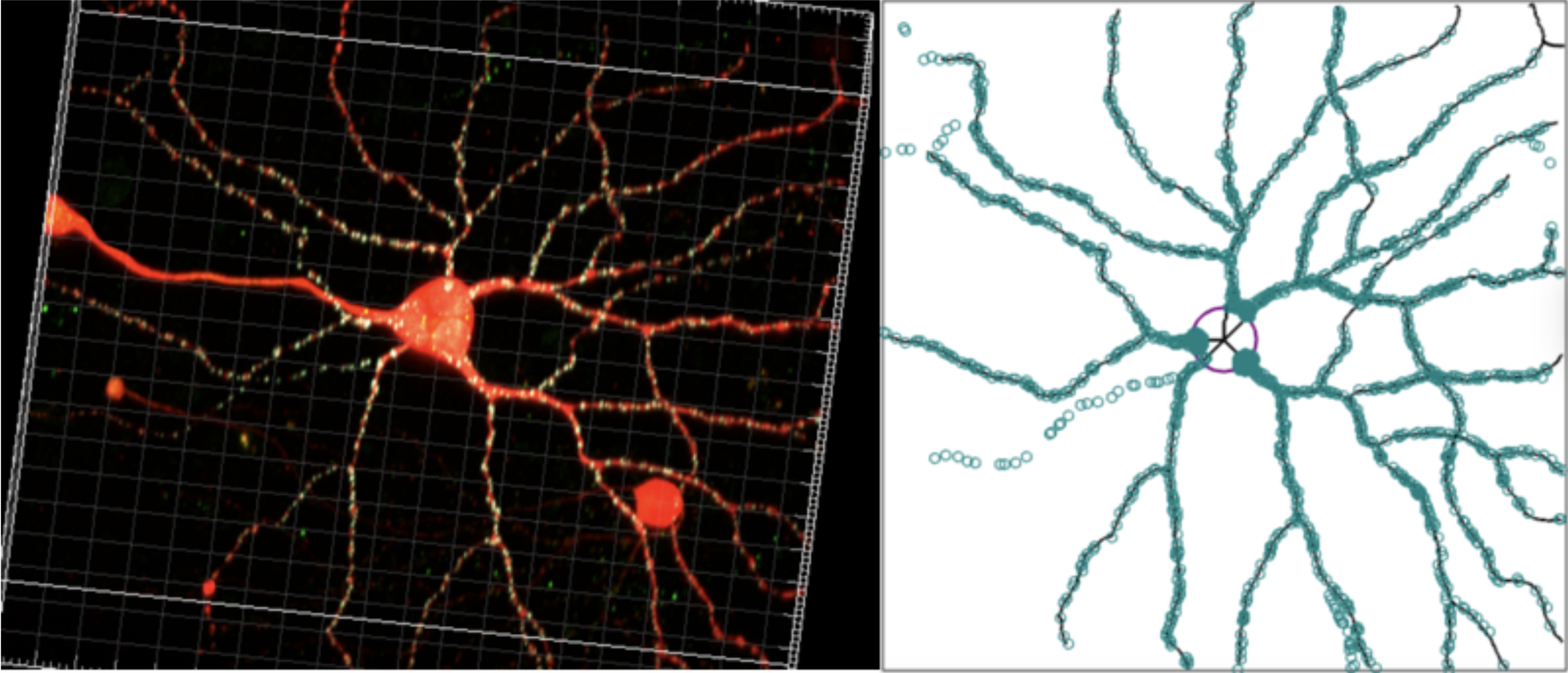
Luca Della Santina, PhD, assistant professor at the University of Houston, provided an example of this approach. He and his colleagues created ObjectFinder, a computational tool for identifying synapses, the tiny points of contact between nerve cells, that is now openly available. He explained: “I don’t have any associated financial benefit from it because I made it open source for everyone. Everyone is encouraged to use it for free and modify it and contribute to the field.”
ObjectFinder: Left: A microscope image of a nerve cell (red) dotted with thousands of synapses (yellow). Right: ObjectFinder traced the branched outline of the cell and identified and registered each synapse (green circles)
An additional argument for resource sharing is grounded in public accountability. Data and resource sharing moves science forward by promoting transparency, by preventing scientists from inadvertently duplicating each other’s work, and by fostering a collaborative research environment.
Two Areas of Glaucoma Research Highlight a Knowledge Gap
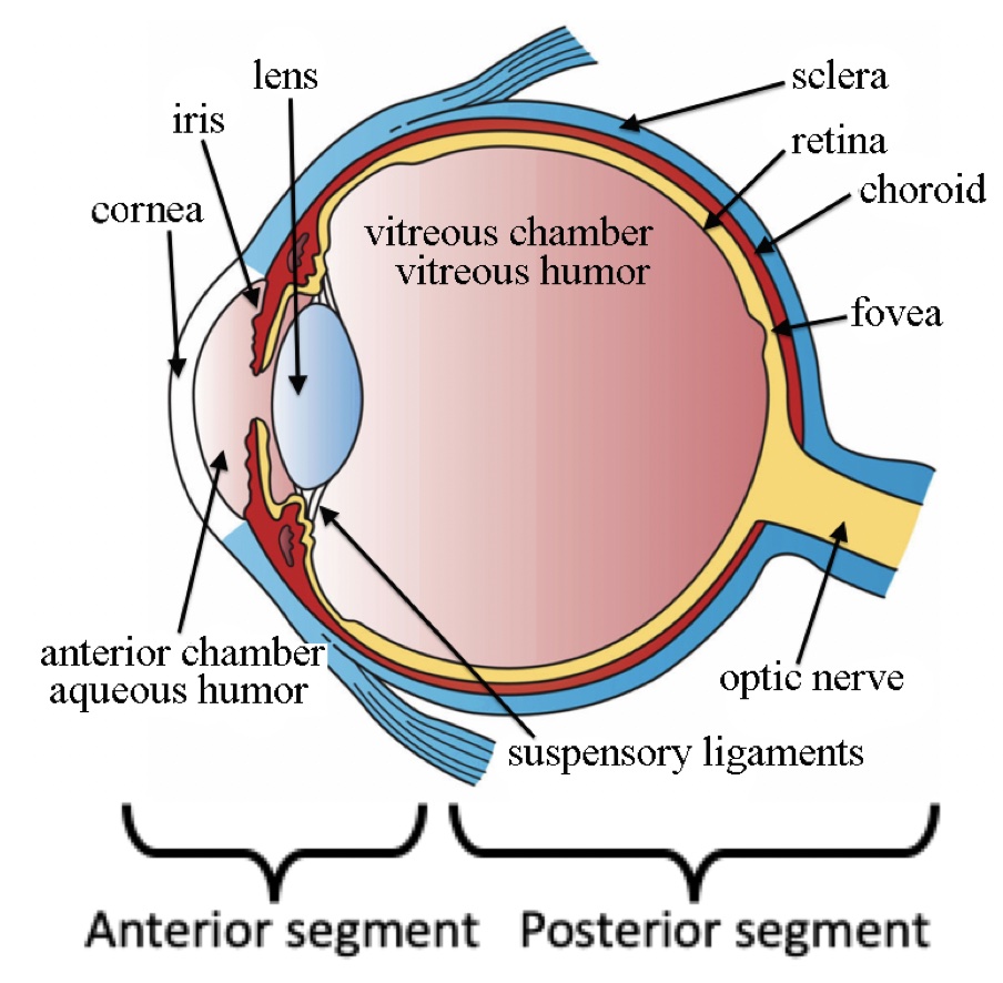
Glaucoma researchers focus on one of two areas: Front-of-the-eye researchers study how fluid is drained to regulate intraocular eye pressure (IOP) and test ways to lower it through medication or surgery. Back-of-the-eye researchers investigate how changes in the retina and the optic nerve correlate with vision loss, the mechanisms of optic nerve damage, and how to protect nerve fibers.
It is still not entirely clear how IOP and optic nerve damage are connected. While it is well established that elevated IOP is the major risk factor for glaucoma, it is not the only cause, and many questions remain, including:
Why do some people have elevated IOP but no glaucoma, while others have glaucoma but no elevated IOP?
Why are some people’s optic nerves more resilient and can withstand elevated IOP better?
Why does pressure reduction help many, but not all patients?
How precisely does elevated IOP damage the optic nerve?
Can the optic nerve be protected even when IOP remains elevated?
Intraocular pressure is regulated by the front of the eye (anterior segment), while the retina and optic nerve are located within the back of the eye (posterior segment). Wikimedia Commons
These unanswered questions highlight the importance of bridging the gap between the front-of-the eye and back-of-the-eye camps to truly understand the disease mechanisms that underlie glaucoma. Significant progress has been made on both fronts, including advanced surgical methods to lower IOP, cutting-edge imaging tools to detect optic nerve damage earlier, and large datasets of clinical data encompassing IOP and optic nerve damage that might identify common elements.
Making the Best Out of Old and New Technologies
Many new tools center around harnessing AI and machine learning to tackle problems such as predicting a patient’s disease course or analyzing images of the retina. Others focus on the power of stem cells that allow researchers to model disease in a dish using a patient’s own cells, with great promise for better individualized treatment options.
Yet sometimes it might be informative to look back into history and learn from our scientific past. Case in point, Dr. Tasneem Sharma, PhD, assistant professor of ophthalmology at Indiana University, provided some fascinating historical background.
Studying the retina dates back nearly 150 years, to the work of Wilhelm Kühne and Julius Steiner, physiologists at the University of Heidelberg, Germany, who pioneered whole-eye cultures of frog eyes. In 1956, Dr. Erich Bauereisen and his colleagues measured the electrical activity in the retinas of isolated frog eyes, figuring out how to keep them alive in a petri dish using the correct temperature and the right amounts of oxygen and glucose. This work helped pave the way for modern-day cell culture.
Fast forward to today, where Dr. Sharma’s colleague at Indiana University and National Glaucoma Research grantee, Jason Meyer, PhD, professor of medical and molecular genetics, introduced his 21st century version of “eyes in a dish.”
It starts with taking a few skin cells from someone – a person affected by glaucoma or a control individual – and treating them with chemicals that eventually transform them into the cells that form the retina. Dr. Meyer then goes one step further and makes what are called “organoids”: retinal cells are grown until they clump together and organize themselves into three-dimensional “mini-retinas in a dish” that are remarkably similar to their real-life counterparts. The cells in these organoids share all of the genetic risk factors with the person from whom they were made, and can be used for example to test an individual’s response to medication or elevated pressure.
In a way, this modern version of Kühne and Steiner’s “eye in a dish” underscores how progress in vision science is driven not only by cutting-edge technologies, but also by an appreciation of the work that laid the foundations for today’s advances.
Glaucoma remains a leading cause of vision impairment, highlighting the need for continued research support. BrightFocus is proud to be a leading funder of glaucoma research worldwide, with research grants totaling $1.8 million funded in 2025 through its National Glaucoma Research program.
The next Glaucoma Fast Track will be held in 2027. Updates will be shared on our website.
Yvonne Ou,MD, University of California, San Francisco: What is Glaucoma? Clinical Aspects of Glaucoma Moderator: Diane Bovenkamp, PhD
Session 1: Fundamentals
Michael Elliott, PhD, University of Oklahoma Health Science Center: Pressure-Induced Models for the Trabecular Meshwork (TM)
Robert W. Nickells, PhD, University of Wisconsin-Madison: Mouse Optic Nerve Crush Model
Rebecca M. Sappington, PhD, Wake Forest University School of Medicine: Pressure-Induced Model
Colleen McDowell, PhD, University of Wisconsin-Madison: Transgenic Models
Moderator: Abbott F. Clark, PhD
Session 2: New Models and Modeling Outcomes
Gillian J. McLellan, BVMS, PhD, University of Wisconsin-Madison: Large Animal Models
Brad Fortune OD, PhD, Devers Eye Institute: Best Ways to Image andSelect Outcomes
Jason Meyer, PhD, Indiana University School of Medicine: Stem Cells
Tasneem P. Sharma, PhD, Indiana University: Perfusion Organ Cultures and the Application of Stem Cells
Moderator: Colleen McDowell, PhD
Session 3: AI and Imaging
Kevin C. Chan, PhD, Stanford University: Overview of Imaging: OCT, MRI, Eye-Brain Connection
Luca Della Santina, PhD, University of Houston: AI Approach to Identify Synapses
Michael G. Anderson, PhD, University of Iowa: AI Approach to Count Axons
Cecilia Lee, MD, Washington University: AI-Powered Insights from Clinical Data: Advancing Glaucoma Research
Moderator: Rebecca M. Sappington, PhD
Session 4: Genetics/Future Therapies
Janey L. Wiggs, MD, PhD, Harvard Medical School: Update on Genetics
Yutao Liu, PhD, Medical College of Georgia: Using GWAS Data in Basic Research
Ahmara G. Ross, PhD, University of Pennsylvania: Challenges of Using Gene Therapy in Glaucoma
Moderator: Abbott F. Clark, PhD
Session 5: Financing Scientific Pursuits/Goals
Paloma Liton, PhD, Duke University
Abbott F. Clark, PhD, University of North Texas Health Science Center
Jimmy Liu, PhD, BrightFocus Foundation
Cynthia Steel, PhD, Glaucoma Research Foundation
Moderator: Rebecca M. Sappington, PhD
Thank you to our sponsors of this year’s Glaucoma Fast Track:
BrightFocus Foundation Board of Directors
RKD Group
J.P. Morgan Private Bank
Atlantic Union Bank
Corentus
Copilevitz, Lam & Raney
Stelter
Moore
About BrightFocus Foundation
BrightFocus Foundation is a premier global nonprofit funder of research to defeat Alzheimer’s, macular degeneration, and glaucoma. Since its inception more than 50 years ago, BrightFocus and its flagship research programs—Alzheimer’s Disease Research, Macular Degeneration Research, and National Glaucoma Research—has awarded more than $300 million in research grants to scientists around the world, catalyzing thousands of scientific breakthroughs, life-enhancing treatments, and diagnostic tools. We also share the latest research findings, expert information, and resources to empower the millions impacted by these devastating diseases. Learn more at brightfocus.org.
Disclaimer: The information provided here is a public service of BrightFocus Foundation and is not intended to constitute medical advice. Please consult your physician for personalized medical, dietary, and/or exercise advice. Any medications or supplements should only be taken under medical supervision. BrightFocus Foundation does not endorse any medical products or therapies.
Share this post
News
Related Articles and Information
Article
National Glaucoma Research Report: Winter 2026
In this issue: Using AI to Detect Glaucoma Earlier, Make the Most of Your Eye Doctor Appointments, Researcher Spotlight: Mengya Zhao, PhD, and more!
Article
How Early Research Funding Is Transforming Glaucoma Detection
If you’ve been diagnosed with glaucoma in the past 15 years, you have likely unknowingly witnessed what early research funding can do for the 4 million Americans living with this sight-stealing disease.
Article
National Glaucoma Research Report: Fall 2025
In this issue: Targeting a Key Protein to Prevent Glaucoma • The Dilated Eye Exam: Your Window to Eye Health • Researcher Spotlight: Shruti Patil, PhD • And More!
Article
Protecting Vision: A New Approach to Scar-Free Healing From Glaucoma Surgery
BrightFocus National Glaucoma Research-funded scientist Jennifer Fan Gaskin, MD, is exploring safer, more effective treatments to prevent scarring after glaucoma filtration surgery, also known as trabeculectomy.
Article
National Glaucoma Research Grantee Receives $2.2 Million Follow-On Award Aimed at Curing Blindness
Additional funding is propelling BrightFocus-funded scientist Jason Meyer, PhD, toward advances in whole-eye transplantation.
Article
Harnessing Artificial Intelligence to Transform Glaucoma Detection
A National Glaucoma Research-funded scientist is pioneering the use of artificial intelligence and telemedicine to detect glaucoma earlier, improve access to care, and prevent irreversible blindness.
Your support helps fund critical research that could prevent vision loss, provide valuable information to the public, and cure this sight-stealing disease.


