A Key Model to Study Cellular Functions to Better Treat Glaucoma
BrightFocus National Glaucoma Research-funded scientist Samuel Herberg, PhD, is using a 3D model of the fluid drainage tissue that could improve our ability to understand and treat glaucoma.
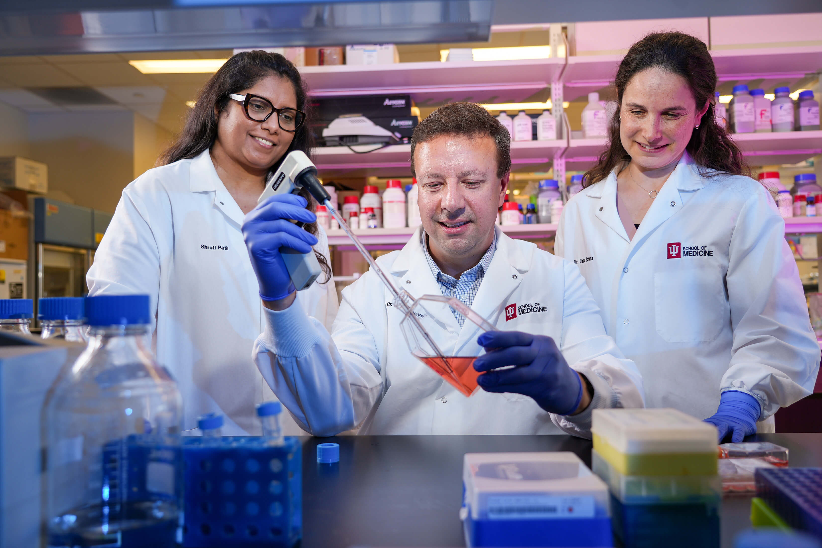
3D models like the one developed by Dr. Samuel Herberg are helping researchers better understand how healthy eye cells function—an essential step toward uncovering what goes wrong in glaucoma and paving the way for new treatments.
Key Takeaways
A team led by BrightFocus National Glaucoma Research grant recipient Samuel Herberg, PhD, is using his unique 3D fluid drainage tissue model to advance our understanding of glaucoma.
Dr. Herberg studies how disease-related changes in the cells’ surroundings affect healthy Schlemm’s canal cells. He focuses on how physical forces—such as pressure, stretching, and stiffness—influence how these cells work, a field known as mechanobiology.
Studies on this 3D model may help develop future treatments for glaucoma.
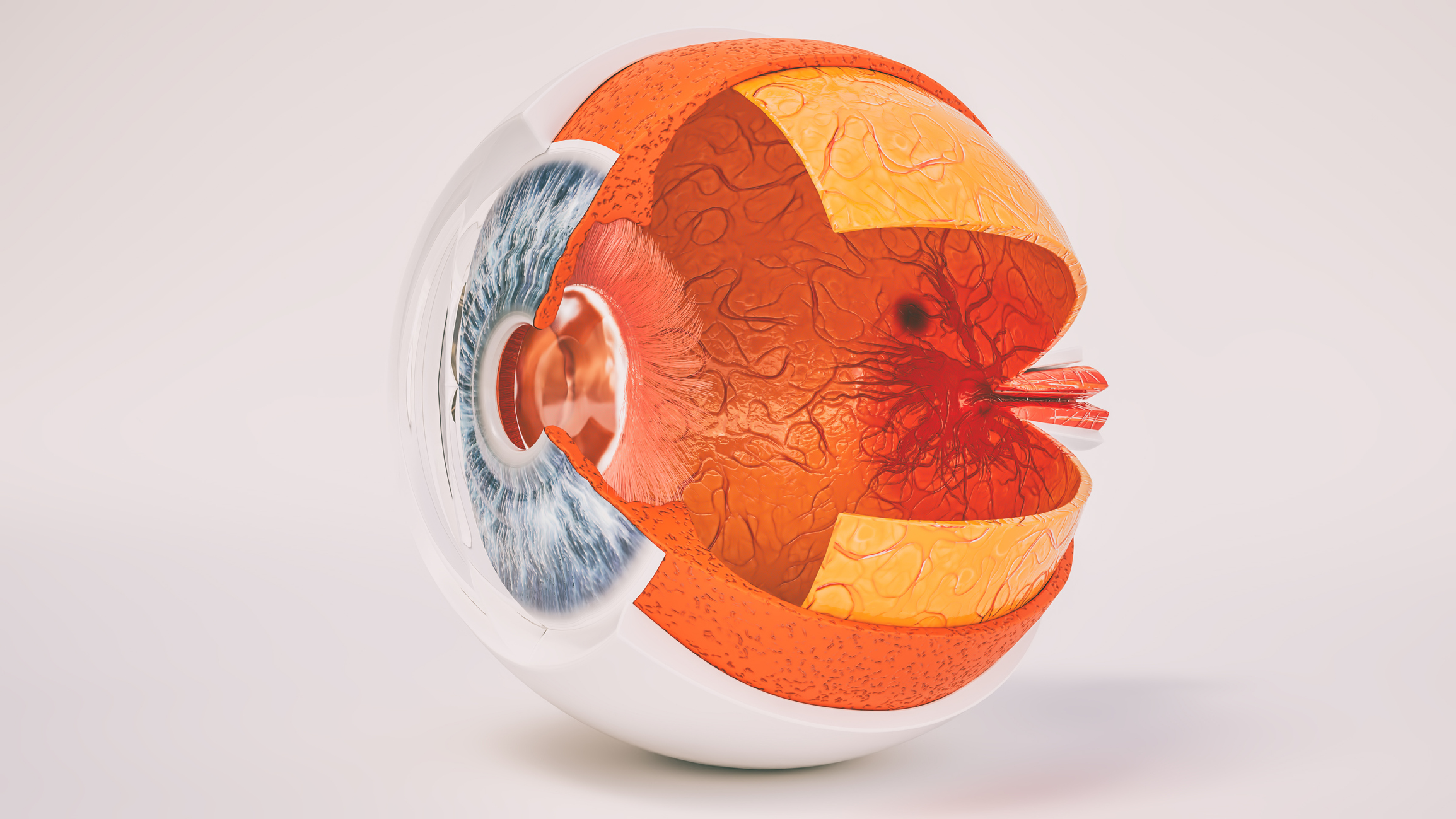
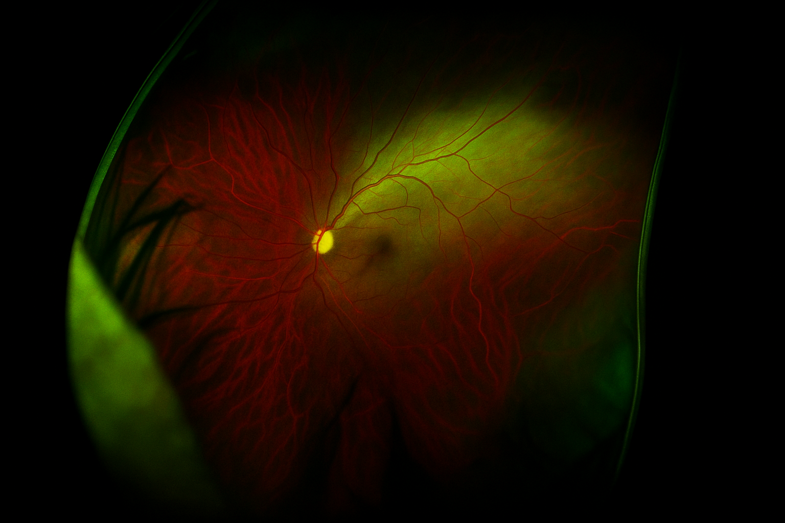
High-pressure glaucoma is the most common form of glaucoma. In general, individuals with increased pressure in the eye are more likely to develop glaucoma. Damage to the optic nerve—the main connection between the eye and the brain—occurs when fluid drainage from the eye fails as glaucoma advances.
Experts don’t fully understand how the cells responsible for fluid transport really function in healthy eyes or what happens within the tissue that causes high pressure to develop in disease. But the damage to the optic nerve is irreversible, and there is no cure to date. New research to better understand how key cells in the eye tissue work could open the door to developing new medicines.
BrightFocus National Glaucoma Research grant recipient Samuel Herberg, PhD, principal investigator at SUNY Upstate Medical University, is using his new research model that mimics the 3D fluid drainage tissue in the front part of the eye to study how the cells behave under high pressure. He believes that understanding the cell behavior in normal conditions could bring us closer to the development of novel treatments for glaucoma.
“The idea of creating a disease model by focusing first on the healthy tissue and learning about the healthy cells and their microenvironment in a way that hasn’t really been done before is where I think we have a unique opportunity [to advance in the glaucoma field],” he said.
Bringing Insights From Bone Healing to Glaucoma
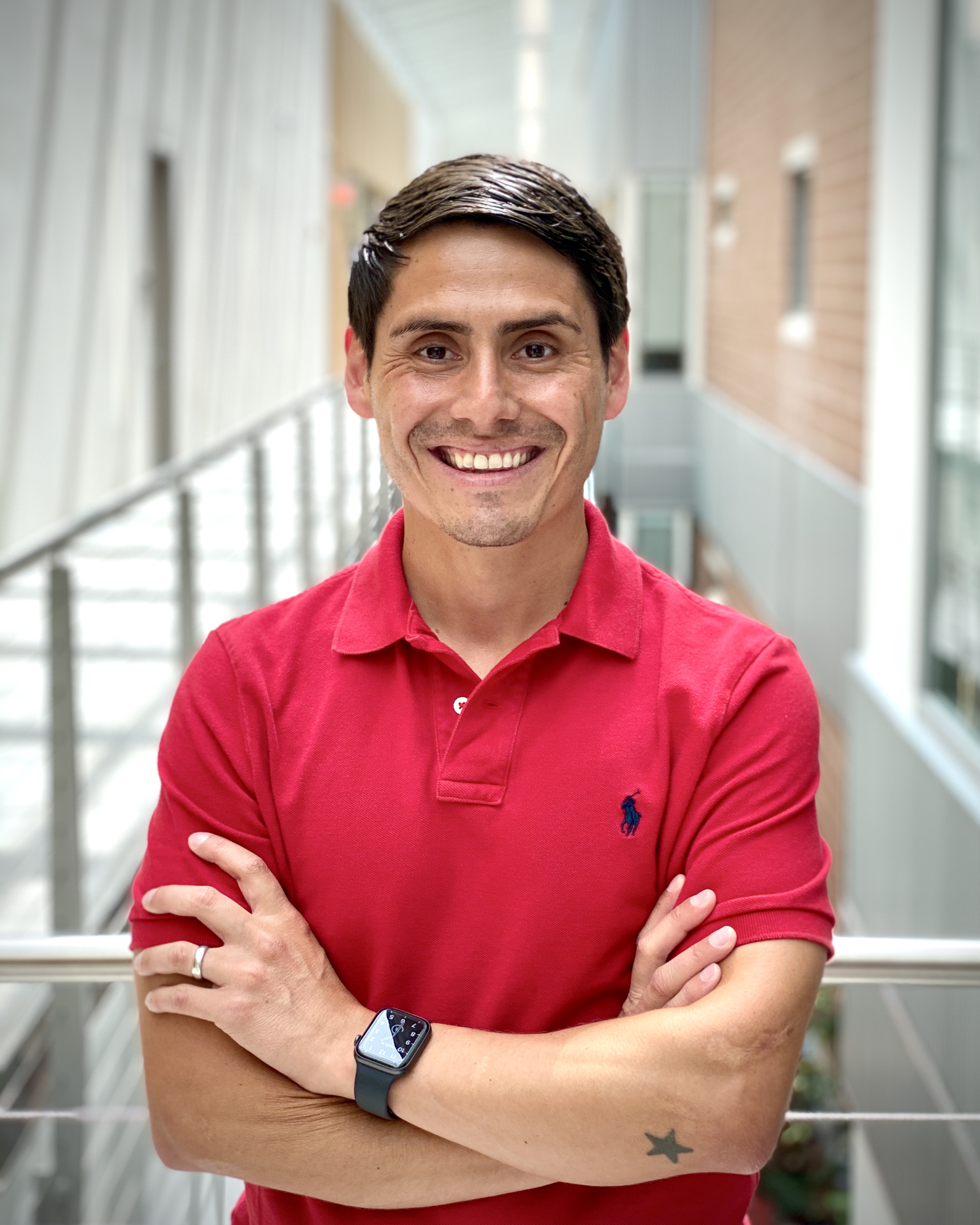
Samuel Herberg, PhD, National Glaucoma Research grant recipient
Under healthy conditions, the eye produces fluid that is constantly draining out of it through the biological drainage system. Fluid flows through a fine, sponge-like structure called the trabecular meshwork, from where it drains into a small channel called Schlemm’s canal. Finally, it enters the body’s veins, like any other waste fluid. But when fluid inside the eye doesn’t drain out the way it should, it builds up, increasing the pressure and ultimately damaging the optic nerve.
Gaining a better understanding of how physical forces, like pressure, stretching, or stiffness, affect the behavior of healthy Schlemm’s canal cells could yield key insights into the mechanobiology of diseased ones. This, in turn, may help identify glaucoma-specific signatures that could serve as targets for treatment.
Dr. Herberg actually started his career in musculoskeletal tissue engineering, where he studied how to guide a special type of cell to become a bone cell. While he was pursuing his doctoral degree, he met his future wife, a vision researcher. Dr. Herberg eventually realized that he could leverage his understanding of tissue engineering in the glaucoma field.
“I happen to have some ideas and tools that were common practice in the musculoskeletal world, but not so widely used yet in the vision world,” said Dr. Herberg. “I felt confident that I could use my technical knowledge and my engineering background to recreate an environment that is instructive to the cells of interest [within the eye].”
One evening in 2018, the couple sat down at the dinner table and brainstormed the idea of working together. They’ve been colleagues since then. While his wife, a clinician-scientist and glaucoma specialist, focuses her research on the optic nerve, Dr. Herberg has taken on the front part of the eye.
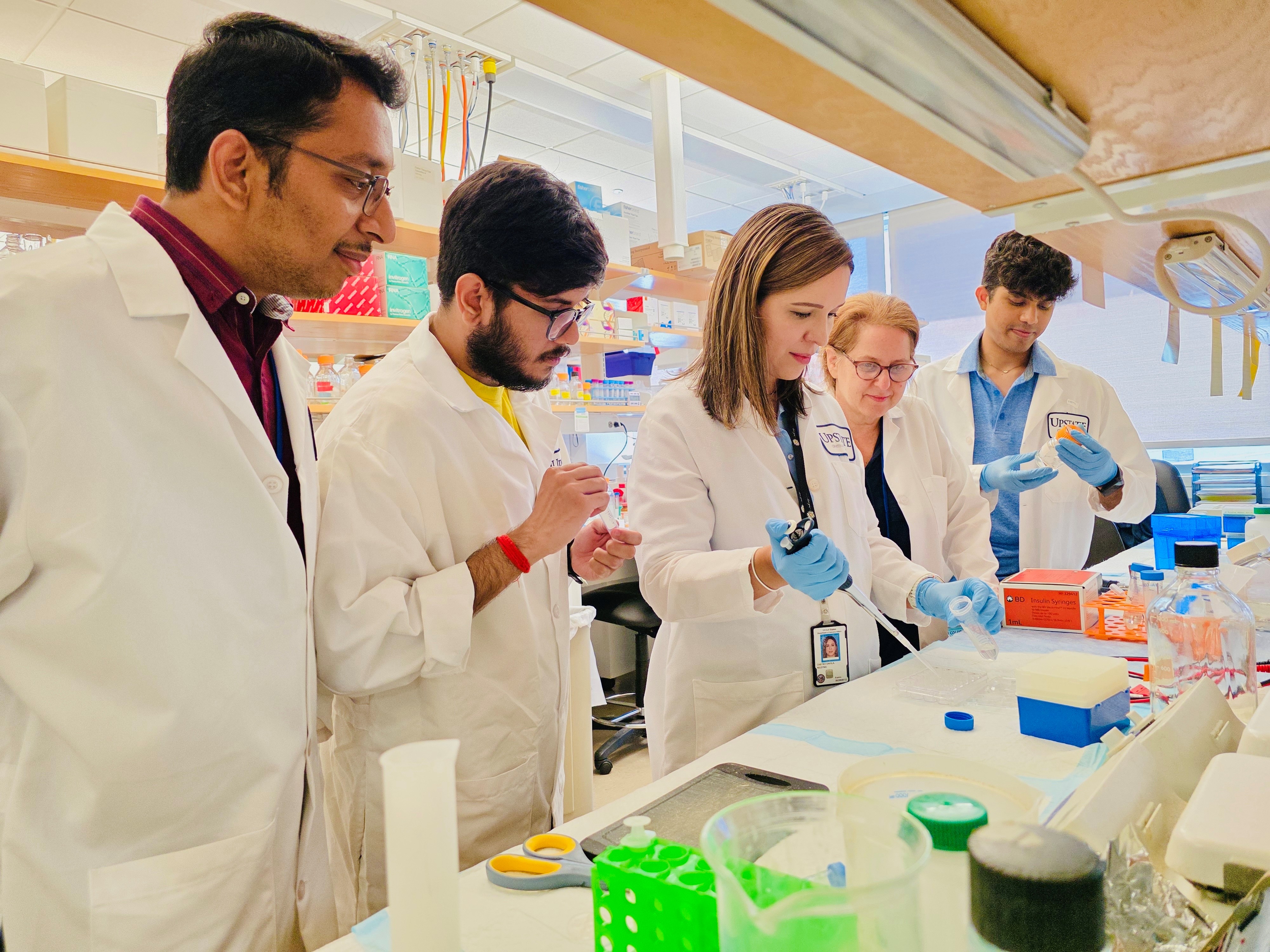
“We take advantage of the joint training environment; my students get to work with my wife, I work with her students,” Dr. Herberg said. “It’s a really dynamic group of trainees.”
Understanding Healthy Eyes to Treat Those With Glaucoma
With support from National Glaucoma Research, Dr. Herberg’s team is using their unique 3D cellular model that simulates the Schlemm’s canal microenvironment in innovative ways. “We use extracellular matrix proteins found in the native tissue and recreate an artificial environment in 3D in which the cells can then live to study their behavior under different conditions,” said Dr. Herberg.
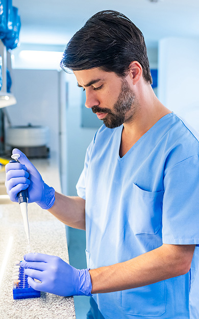
The researchers rely on donor eye tissue from healthy individuals and people living with glaucoma to isolate both the trabecular meshwork and Schlemm’s canal cells for their study. “We wouldn’t be able to do what we do without tissue donors,” Dr. Herberg said. “I’m immensely grateful for their gift.”
Dr. Herberg noted that until now, there were no accurate models to properly study Schlemm’s canal cell mechanobiology in the lab. He said that when he’s using his model, “some of the cell behaviors are much more similar to the native tissue compared to conventional culture,” highlighting how important the development of this unique 3D model was to reflect real-life biology more accurately. “It is the closest approximation to the actual tissue we can build reliably in the laboratory setting,” he added.
In today’s challenging funding climate, Dr. Herberg believes his research into the biological mechanisms of glaucoma simply wouldn’t have gotten off the ground without the support from BrightFocus programs, particularly those focusing on the cellular mechanisms of glaucoma. “I’m immensely grateful for alternative ways of pursuing vision research in glaucoma through National Glaucoma Research,” he said.
“I’m immensely grateful for alternative ways of pursuing vision research in glaucoma through National Glaucoma Research.”
About BrightFocus Foundation
BrightFocus Foundation is a premier global nonprofit funder of research to defeat Alzheimer’s, macular degeneration, and glaucoma. Since its inception more than 50 years ago, BrightFocus and its flagship research programs—Alzheimer’s Disease Research, Macular Degeneration Research, and National Glaucoma Research—has awarded more than $300 million in research grants to scientists around the world, catalyzing thousands of scientific breakthroughs, life-enhancing treatments, and diagnostic tools. We also share the latest research findings, expert information, and resources to empower the millions impacted by these devastating diseases. Learn more at brightfocus.org.
Disclaimer: The information provided here is a public service of BrightFocus Foundation and is not intended to constitute medical advice. Please consult your physician for personalized medical, dietary, and/or exercise advice. Any medications or supplements should only be taken under medical supervision. BrightFocus Foundation does not endorse any medical products or therapies.
Protecting Vision: A New Approach to Scar-Free Healing From Glaucoma Surgery
BrightFocus National Glaucoma Research-funded scientist Jennifer Fan Gaskin, MD, is exploring safer, more effective treatments to prevent scarring after glaucoma filtration surgery, also known as trabeculectomy.
Article
National Glaucoma Research Grantee Receives $2.2 Million Follow-On Award Aimed at Curing Blindness
Additional funding is propelling BrightFocus-funded scientist Jason Meyer, PhD, toward advances in whole-eye transplantation.
Article
Harnessing Artificial Intelligence to Transform Glaucoma Detection
A National Glaucoma Research-funded scientist is pioneering the use of artificial intelligence and telemedicine to detect glaucoma earlier, improve access to care, and prevent irreversible blindness.
Article
How Some Eye Cells Survive Glaucoma
BrightFocus National Glaucoma Research-funded scientist Mengya Zhao, PhD, uses a unique combination of techniques to understand why certain retinal cells stay strong in glaucoma while others don’t.
Article
How the Breakdown of Cellular ‘Powerhouses’ Drives Exfoliation Glaucoma
A study yields new insights that could inspire novel treatments for exfoliation glaucoma.
Article
Looking Beyond Pressure-Lowering Drugs to Treat Glaucoma
A National Glaucoma Research-funded scientist is building a 3D model of the optic nerve that she hopes will solve unanswered questions about glaucoma—and inspire new treatments.
Your support helps fund critical research that could prevent vision loss, provide valuable information to the public, and cure this sight-stealing disease.