

Exfoliation Syndrome and Lysosomal Disease

About the Research Project
Program
Award Type
Standard
Award Amount
$50,000
Active Dates
July 01, 2015 - June 30, 2016
Grant ID
G2015195
Co-Principal Investigator(s)
J Wolosin, PhD, Icahn School of Medicine at Mount Sinai
Goals
In exfoliation syndrome (XFS), the eye starts accumulating protein aggregates that resemble fluffy white deposits. Eventually this debris may block the exit of fluid from the eye, causing a buildup of pressure that is believed to contribute to glaucoma and optic nerve damage. If unchecked, these changes can lead to blindness. We have discovered that cells obtained from XFS eyes may have a problem degrading these protein aggregates, leading to the toxic accumulation of this form of “cellular trash.” Our project will test methods to accelerate degradation of this cellular waste in order to improve the health of XFS-affected eyes.
Summary
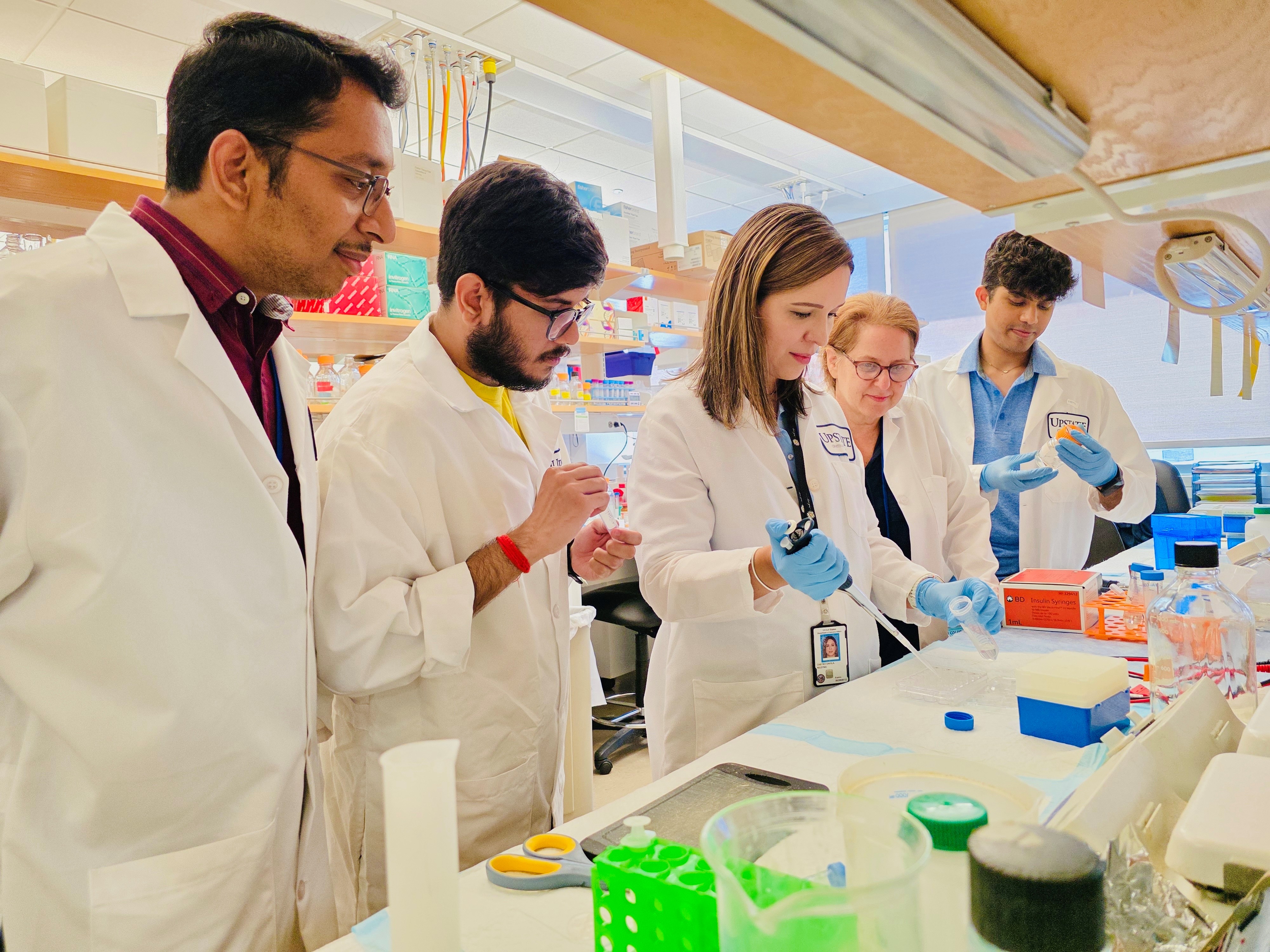
Our goal is to reverse the effects of exfoliation syndrome (XFS), the leading identifiable cause of glaucoma. Our lab is investing the hypothesis that eyes with XFS have a dysfunction in the cellular machinery that would normally degrade these protein aggregates, causing the accumulation of cellular waste that becomes toxic. We are growing cells from tissue derived from XFS patients who have undergone surgery to have the exfoliated tissue removed from their eyes to relieve pressure. These cells are called tenon fibroblasts. We are using the tenon fibroblasts as a model system to investigate methods to accelerate degradation of cellular waste in order to improve the health of XFS-affected cells.
Inside the cell, lysosomes degrade cellular waste and mitochondria produce the energy to power the cell. Our work links XFS in the eye to lysosomal and mitochondrial age-related malfunction and disease. Recent work in studies of several other age-related diseases, such as age-related macular degeneration (AMD) and Alzheimer’s disease, have found that lysosomal and mitochondrial dysfunction are significant contributors to disease pathology. Since one of the major challenges of XFS research has been a lack of model systems for experimental purposes, our patient-derived cells provide a wealth of opportunity to identify critical pathways that are disrupted in XFS-affected cells.
Any evidence we are able to demonstrate in support of this central hypothesis will elucidate cellular mechanisms particular to XFS and reveal novel approaches that prevent XFS manifestation in the eye. The correlation with other age-related diseases opens the door to new therapies for XFS, some of which may already be under development.
Related Grants
National Glaucoma Research
Maldevelopment of Schlemm's Canal in Children with Glaucoma
Active Dates
July 01, 2026 - June 30, 2028

Principal Investigator
Revathi Balasubramanian, PhD
Current Organization
Columbia University
Maldevelopment of Schlemm's Canal in Children with Glaucoma
Active Dates
July 01, 2026 - June 30, 2028
Principal Investigator
Revathi Balasubramanian, PhD
Current Organization
Columbia University
National Glaucoma Research
The Immune Landscape of the Human Optic Nerve Head
Active Dates
July 01, 2026 - June 30, 2028

Principal Investigator
Monica Vetter, PhD
Current Organization
University of Utah
The Immune Landscape of the Human Optic Nerve Head
Active Dates
July 01, 2026 - June 30, 2028
Principal Investigator
Monica Vetter, PhD
Current Organization
University of Utah
National Glaucoma Research
Understanding Optic Nerve Head Astrocyte Contributions to Glaucoma
Active Dates
July 01, 2026 - June 30, 2028

Principal Investigator
Evan Cameron, PhD
Current Organization
Cleveland Clinic - Lerner Research Institute
Understanding Optic Nerve Head Astrocyte Contributions to Glaucoma
Active Dates
July 01, 2026 - June 30, 2028
Principal Investigator
Evan Cameron, PhD
Current Organization
Cleveland Clinic - Lerner Research Institute