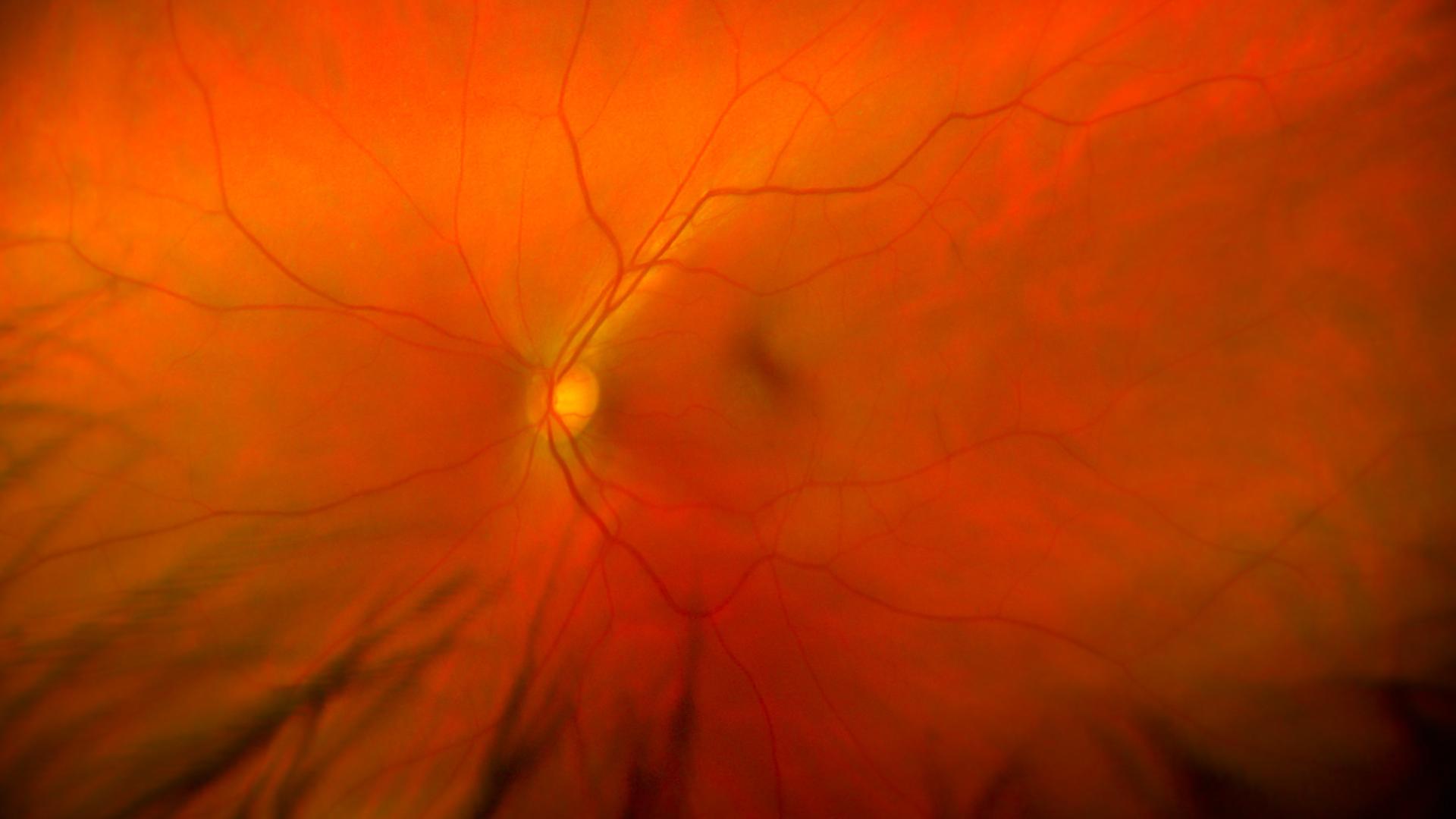
View inside of the human eye showing a healthy retina, optic nerve, and macula.
An international research team funded by BrightFocus Foundation’s Alzheimer’s Disease Research program has successfully developed a first-of-its-kind artificial intelligence (AI) model that could eventually detect Alzheimer’s disease through photographs of the retina, the back of the eye.
This new method would allow people to receive their results immediately and benefit from early intervention. With an accuracy of more than 80%, the screening opens doors to earlier diagnosis and treatment, which are key to slowing the progression of the disease.
A successful AI model
The researchers developed a deep learning AI algorithm to study and compare minute details within the retinal photos, also called fundus photos. Deep learning is a method of teaching computers how to process data in a similar way to how humans learn. The model can recognize complex patterns in photographs and other data to produce accurate results much faster than humans could.
The researchers used nearly 13,000 retinal photographs from 648 people with Alzheimer’s and 3,240 others who did not have the disease. The photographs came from 11 previous studies from different countries, allowing for a diverse patient sample.
The team was led by BrightFocus grantee Carol Cheung, PhD, and conducted at the Chinese University of Hong Kong’s Faculty of Medicine. Details of the research were published in The Lancet Digital Health.
“Our AI model retained a robust ability to differentiate between subjects with and without Alzheimer’s disease, even in the presence of concomitant eye diseases like macular degeneration and glaucoma,” Dr. Cheung said in a statement from the university. “This further supports the concept that our AI analysis of fundus photographs is an excellent tool for the detection of the memory-depriving Alzheimer’s disease.”
Improving diagnosis
Detecting Alzheimer’s disease in its early stages is key, so that treatment can be started as soon as possible to slow its progression. But early diagnosis relies on a series of cognitive tests, assessments, neuroimaging studies, and cerebrospinal fluid studies. Patients usually are diagnosed only after there has been significant brain degeneration and cognitive decline.
The algorithm developed by the research team provides results immediately, which can enable patients to be treated sooner. The researchers said it’s their hope that in the near future, routine eye exams to look for diseases like glaucoma could include screenings for Alzheimer’s disease as well.
A window to the inner body
The retina is considered a window to the central nervous system because it is the only human tissue allowing direct, noninvasive pictures of degenerative changes in the blood vessels and nerves of the brain. Many health conditions can be identified this way, including cardiovascular disease, Parkinson’s disease, and multiple sclerosis.
The retinal fundus is the interior of the eyeball that includes the light-sensitive retina, the head of the optic nerve, and the part of the eye that produces clear vision, called the macula. Based on this research, retinal fundus photography, which is widely accessible and cost-effective, has the potential to be an important tool for screening people at risk of Alzheimer’s disease.
“In the entire central nervous system, only the blood vessels and nerves in the retina allow direct visualization and analysis,” Dr. Cheung’s fellow researcher, Dr. Clement Tham Chee-yung, said in the same university statement. “Through noninvasive fundus photography, we can detect a range of changes in the blood vessels and nerves of the retina that are associated with Alzheimer’s disease.”
The staggering toll of Alzheimer’s
Alzheimer’s has been described as a thieving disease because it steals the minds of the people it affects. It leads to memory loss, confusion, and an inability to communicate or care for oneself. The disease also takes a toll on family members, who provide the majority of caregiving.
The number of Alzheimer’s cases is expected to triple by the year 2050, an epidemic that could overwhelm the healthcare system. In 2023, Alzheimer’s and other dementias are expected to cost the U.S. $345 billion—a cost that could rise to nearly $1 trillion in 2050.
About BrightFocus Foundation
BrightFocus Foundation is a premier global nonprofit funder of research to defeat Alzheimer’s, macular degeneration, and glaucoma. Since its inception more than 50 years ago, BrightFocus and its flagship research programs—Alzheimer’s Disease Research, Macular Degeneration Research, and National Glaucoma Research—has awarded more than $330 million in research grants to scientists around the world, catalyzing thousands of scientific breakthroughs, life-enhancing treatments, and diagnostic tools. We also share the latest research findings, expert information, and resources to empower the millions impacted by these devastating diseases. Learn more at brightfocus.org.
Disclaimer: The information provided here is a public service of BrightFocus Foundation and is not intended to constitute medical advice. Please consult your physician for personalized medical, dietary, and/or exercise advice. Any medications or supplements should only be taken under medical supervision. BrightFocus Foundation does not endorse any medical products or therapies.
New Blood Test Could Personalize Alzheimer's Treatment Like Never Before
A new Alzheimer’s blood test may reveal how far the disease has progressed and identify who’s most likely to benefit from anti-amyloid therapies.
Article
A BrightFocus-Funded Non-Invasive Treatment Slows Alzheimer's Progression in Yearlong Study
Sinaptica Therapeutics unveils breakthrough Phase 2 results for an experimental non-invasive treatment for Alzheimer’s disease.
Article
Why APOE4 Makes Women More Susceptible to Alzheimer's
The Alzheimer’s risk gene APOE4 puts women at greater risk for Alzheimer’s disease than men. New research unveils a possible cause.
Article
BrightFocus-Supported C2N Diagnostics Receives Transformative Follow-On Funding to Expand Access to Alzheimer's Blood Test
The company behind the first-of-its-kind blood-based screening test for Alzheimer’s disease—rooted in pivotal early BrightFocus support—has received a $15 million investment.
Article
New Study Finds Changes to Blood Immune Cells in Alzheimer's Disease
Immune cells undergo non-inherited genetic changes that are associated with higher Alzheimer’s risk.
Article
Researcher Discovers a Potential Way to Repair Synapses Damaged in Alzheimer's Disease, Restore Memory
A new study asks the question: what if we could reverse the damage caused by Alzheimer’s disease-related proteins like tau? Learn more.
Every Donation is a Step Forward in the Fight Against Alzheimer’s
Your donation powers cutting-edge research and helps scientists explore new treatments. Help bring us closer to a cure and provide valuable information to the public.