Alzheimer’s disease is a progressive neurodegenerative disorder that profoundly impacts brain health, leading to cognitive decline and memory loss. The effects of this disease extend beyond the bounds of normal aging and cause significant damage to the brain.
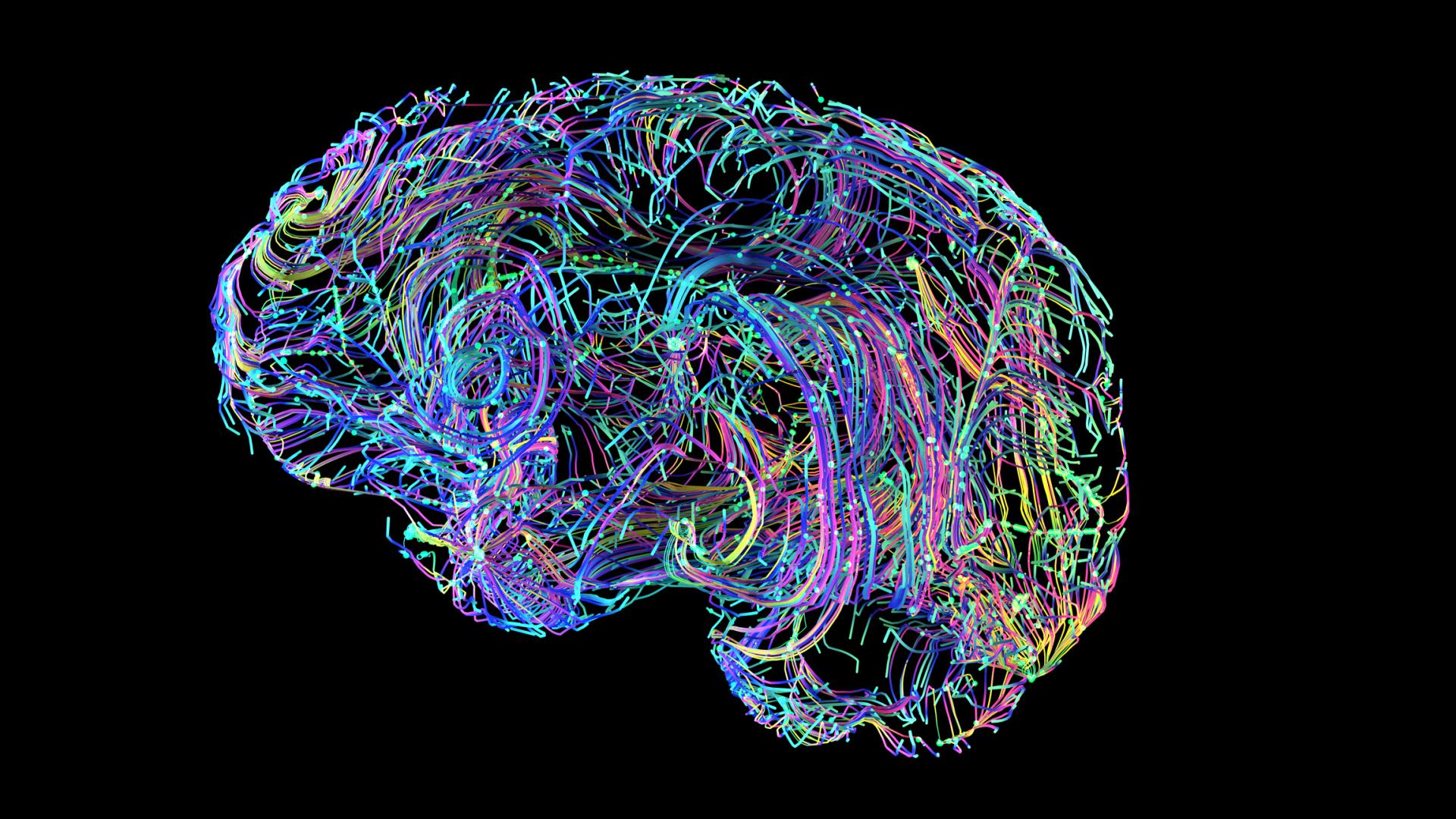
The effects of Alzheimer’s can be seen at every scale—from the tiniest brain cell to the entirety of the brain. To understand how Alzheimer’s affects the brain, it’s helpful to first review the different types of brain cells. They can be broadly categorized based on the roles of communicators, facilitators, janitors, and first responders:
The Communicators—Neurons: These nerve cells underlie the most basic to the most complex activities of daily life. Neurons create telephone chains that can share messages throughout one brain area and across the many regions of the brain.
The Facilitators—Oligodendrocytes: Provide neurons with a specialized fatty insulation so they can share messages faster.
The Janitors—Astrocytes: As the guardians of brain balance, astrocytes help get rid of waste and maintain a harmonious environment. They also support their fellow brain cells, neurons and microglia, through additional activities in damage response, resource recycling, and more.
The First Responders—Microglia: As the immune system of the brain, microglia respond to any cell damage or foreign invaders that may appear in the environment. They can release chemicals that promote cell healing and those that are a key part of immune response.
The normal process of aging and age-related diseases can impact each of these cells and present challenges to brain health. Below, learn some of the many ways aging and Alzheimer’s can affect the brain, and meet a few of the scientists funded by BrightFocus’ Alzheimer’s Disease Research program using this knowledge to fight back.
A Shrinking Brain
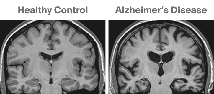
The brain experiences some shrinking with normal aging and significantly more in age-related diseases. With Alzheimer’s, a significant amount of shrinkage occurs because of damage and resulting degeneration from the disease. Alzheimer’s actively attacks brain cells, impairing their function and eventually leading to cell death.
An MRI scan of a non-demented brain and a brain with Alzheimer’s disease. Significant brain shrinkage is observed with the abundance of black space present in the Alzheimer’s image. Photo credit: Neurotorium.org
BrightFocus Alzheimer’s Disease Research grantee, Dr. Zahra Shirzadi, is identifying imaging patterns for injury to the white parts of the brain (shown above) and evaluating their use for predicting Alzheimer’s risk. Learn more.
Buildup of Amyloid-Beta and Tau
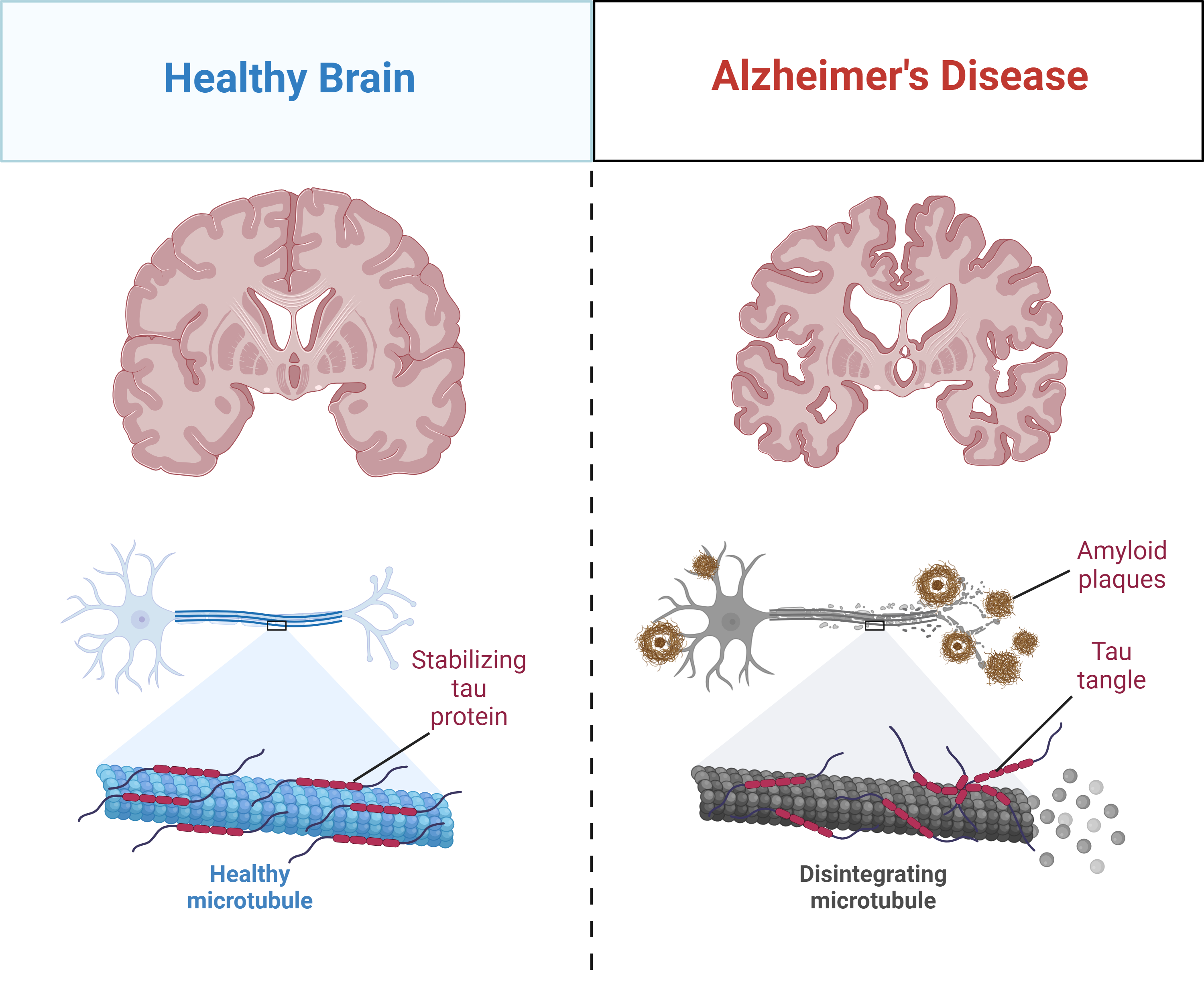
Dr. Alois Alzheimer first discovered Alzheimer’s after observing dense plaques and tangle-like structures in the brain of a middle-aged patient who had experienced memory loss and other symptoms. Plaques and tangles do appear in the brains of elderly, non-demented individuals—but to observe this pathology in a younger patient was peculiar. Scientists would later come to find that these Alzheimer’s hallmarks are the end-product from a decades-long buildup of two proteins: amyloid-beta (plaques) and tau (tangles). Smaller forms of these proteins, called oligomers, may begin damaging the brain long before any symptoms appear.
Brain shrinkage results from progressive damage to brain cells in Alzheimer’s. A healthy neuron (blue; left) can be divided into three areas: a messaging-receiving end (left), a long hallway (middle; “axon”), and a message-sending end (right). Inside the axon is a skeleton of tybe-like structures called microtubules that create a shuttle system between the two ends. Tau stabilizes this structure but becomes misformed and damages the shuttle system in Alzheimer’s (right). Amyloid-beta plaques also surround the neuron, impairing communication and causing additional damage to the cell. Created with BioRender.com
Scientists at C2N Diagnostics used their scientific understanding of amyloid-beta and tau in Alzheimer’s to create a first-of-its-kind blood test with early support from Alzheimer’s Disease Research program. Learn more.
Inflammation Unleashed
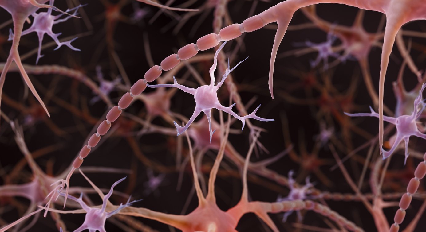
Inflammation is a core part of the immune system response for the brain and body. With age, low-grade chronic inflammation can develop—a phenomenon scientists call inflamm-aging. This process can make the brain more vulnerable to age-related diseases, especially if there is a history of other inflammatory events like a traumatic brain injury. Alzheimer’s-related damage to brain cells causes a significant increase in inflammation— a double-edged sword that can cause further damage.
Pictured here are microglia (pink) floating among neurons (red). Not included in this image are astrocytes, which can also contribute to inflammation.
Alzheimer’s Disease Research grantee Dr. David Gate is investigating how immune cells in the blood and an individual’s environment can contribute to inflammation in Alzheimer’s. Learn more.
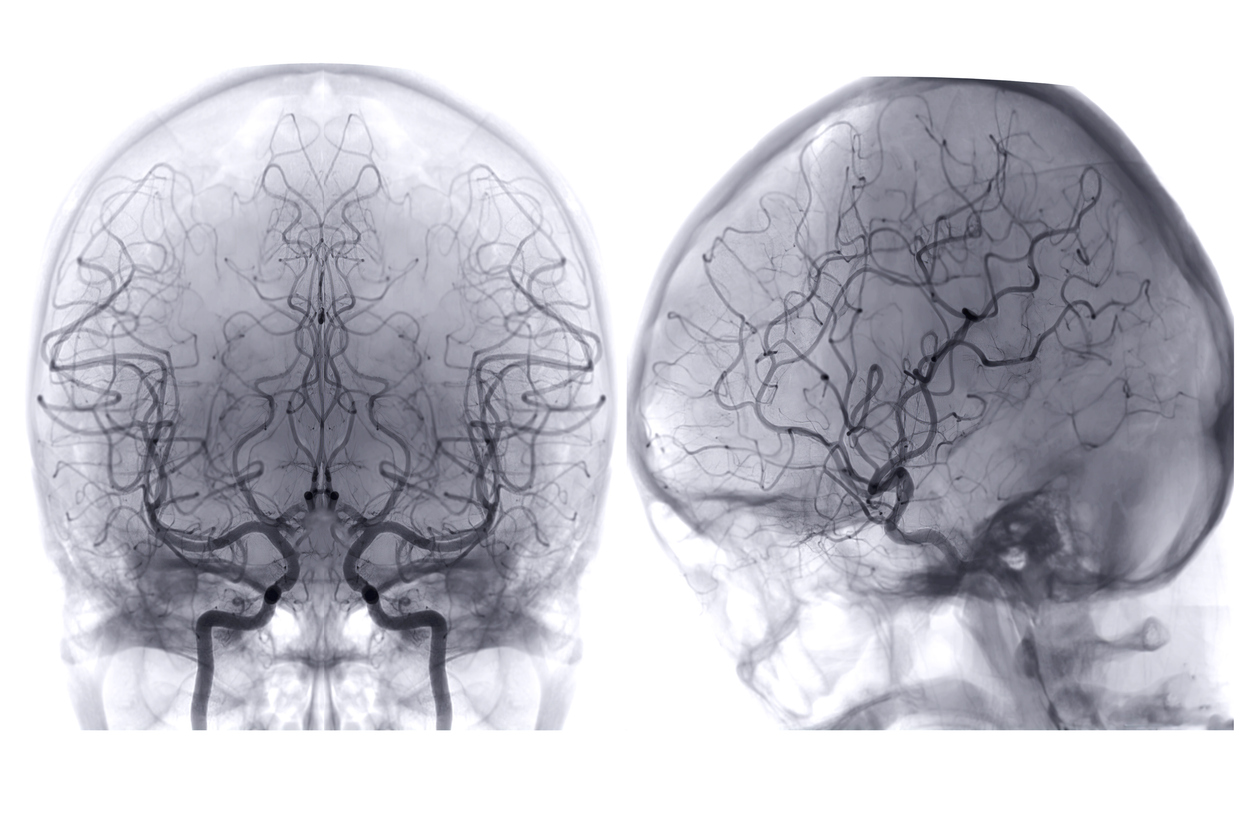
A Loss of Proper Brain Blood Flow
Blood carries essential components to the brain, like oxygen and sugar (glucose), providing energy to support brain activity. Brain blood flow decreases normally with age, and even more with Alzheimer’s. The decline seen in Alzheimer’s can significantly impact brain activity—making even the most basic cell functions difficult. This can have wide-ranging implications for cells and overall brain health and likely contributes to cognitive decline.
Brain blood vessels are visualized through a procedure called cerebral angiography, using a specialized dye and X-rays to observe blood flow.
Alzheimer’s Disease Research grantee Dr. Marta Casquero-Veiga is developing new imaging tools to identify small brain blood clots that develop in Alzheimer’s. Learn more.
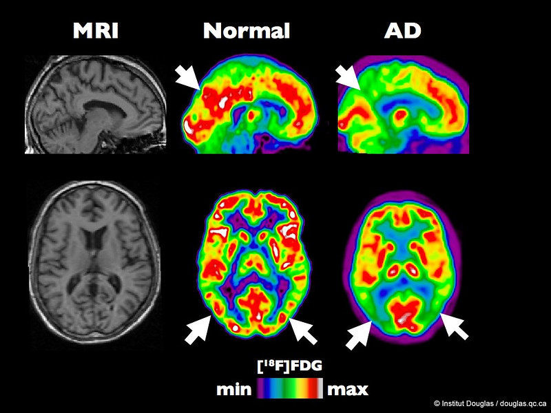
Running Low on Brain Fuel
Glucose (blood sugar) is the main energy source for the brain. Brain cells take up glucose and use it to power their various specialized activities. In Alzheimer’s, the brain becomes deprived of glucose, keeping the cells from doing their jobs. The resulting decrease in brain activity can have consequences for important functions like memory. A loss of balance in glucose throughout the body may also increase the risk for Alzheimer’s, as seen in people with obesity or Type-2 diabetes. Studies are also ongoing to determine if the changes in brain glucose observed in Alzheimer’s are linked to cardiovascular risk factors.
Picture above is a PET scan of a non-demented person and a person with early Alzheimer’s. White arrows point to areas that typically use a lot of energy (middle, red) but lack the same energy in the Alzheimer’s brain (right, green). Credit: Institut Douglas Flickr
With grant funding from Alzheimer’s Disease Research, Dr. Na Zhao investigated how an Alzheimer’s risk gene impairs brain health and looked to insulin as a potential treatment. Learn more.
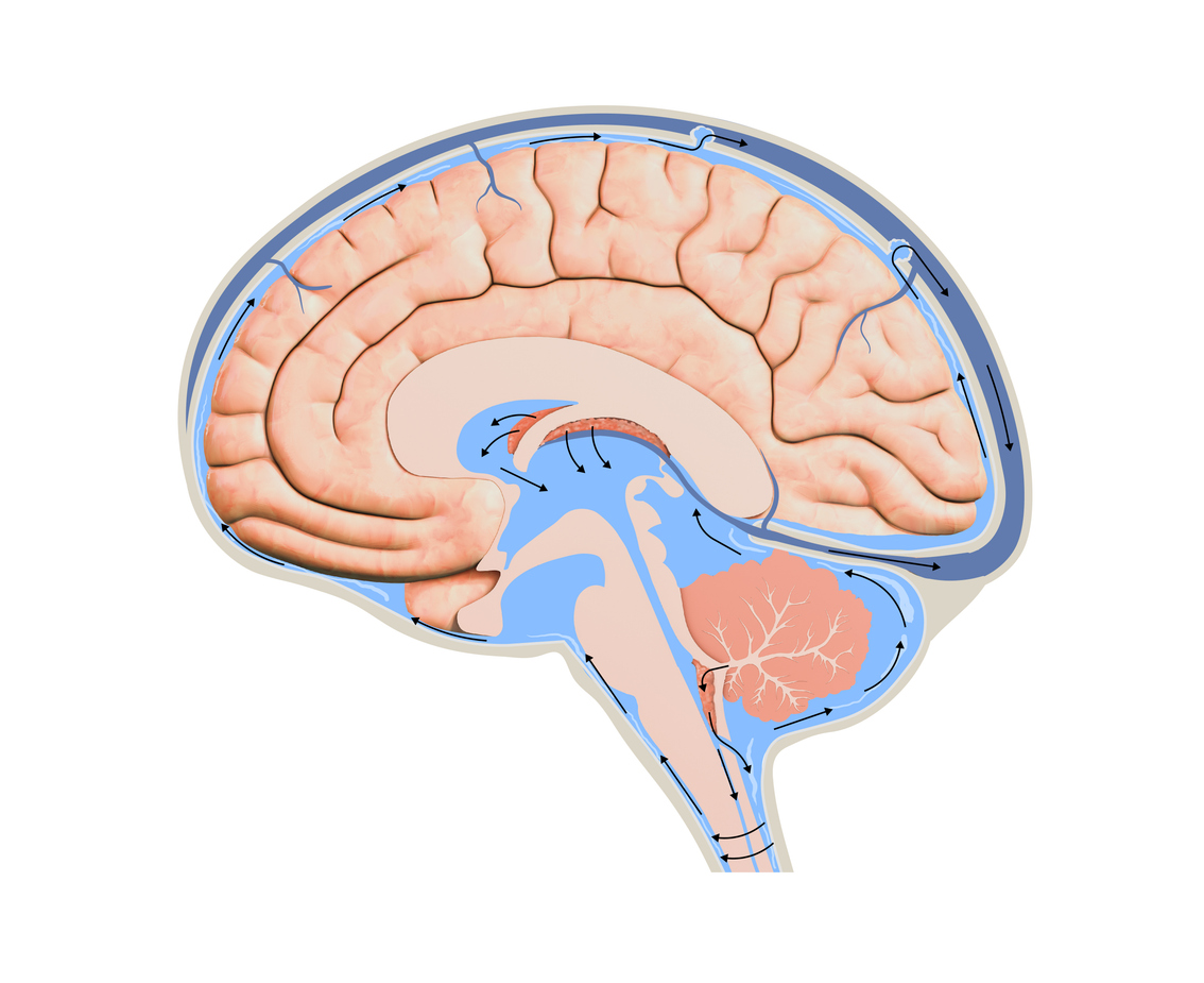
Waste Removal
During sleep, the brain flushes its waste by circulating a substance called cerebrospinal fluid. The clearance of waste from the brain is crucial to maintaining a harmonious environment and preventing disease. With age, normal sleep patterns can become disturbed, leading to a host of consequences for the brain and body. Among them is the potential for proteins, like amyloid-beta and tau, to buildup and perpetuate disease. People with Alzheimer’s can experience sleep disturbances years before the onset of clinical symptoms. Studies are ongoing to determine individual factors that can influence waste removal in Alzheimer’s. Conversely, some research suggests that better waste removal might be tied to cognitive resilience and successful aging.
Cerebrospinal fluid flows throughout and around the brain in a process called lymphatic clearance, meant to rid the brain of waste during sleep.
Dr. Christopher Morrone is using his funding from Alzheimer’s Disease Research to study how a lack of sleep leads to protein buildup in the brain. Learn more.
Summary
Alzheimer’s takes the consequences of normal aging and turns up the intensity to an unbearable level for the brain. Many of the brain’s basic mechanisms like first aid, getting enough fuel, removing waste, and more are turned on themselves and instead, contribute to disease progression.
By understanding the ins-and-outs of Alzheimer’s, researchers can design new detection strategies and treatments. BrightFocus’ Alzheimer’s Disease Research program supports these scientists in propelling next-generation science toward a cure for Alzheimer’s. Discover how BrightFocus is driving innovation in diagnosis and treatment here.
About BrightFocus Foundation
BrightFocus Foundation is a premier global nonprofit funder of research to defeat Alzheimer’s, macular degeneration, and glaucoma. Since its inception more than 50 years ago, BrightFocus and its flagship research programs—Alzheimer’s Disease Research, Macular Degeneration Research, and National Glaucoma Research—has awarded more than $330 million in research grants to scientists around the world, catalyzing thousands of scientific breakthroughs, life-enhancing treatments, and diagnostic tools. We also share the latest research findings, expert information, and resources to empower the millions impacted by these devastating diseases. Learn more at brightfocus.org.
Disclaimer: The information provided here is a public service of BrightFocus Foundation and is not intended to constitute medical advice. Please consult your physician for personalized medical, dietary, and/or exercise advice. Any medications or supplements should only be taken under medical supervision. BrightFocus Foundation does not endorse any medical products or therapies.
Brain Health
Share this post
Resources
Related Articles and Information
Expert Information
Expanding the Alzheimer's Treatment Landscape: A 2026 Forecast
Learn about the treatments landscape for Alzheimer’s in 2026 and beyond.
Expert Information
Alzheimer's Risk Reduction: Nutrition & Lifestyle
There are healthy actions people can take to improve and maintain health, no matter what conditions they may be facing.
Recursos en español
Recursos útiles para el cuidado del Alzheimer y la demencia
Una lista de recursos de la enfermedad de Alzheimer para pacientes y cuidadores.
Expert Information
Why Some Brains Stay Sharp With Age
Learn how cognitive reserve can make some people more resistant to memory decline.
Downloadable Resource
Understanding Alzheimer’s Disease
Alzheimer’s disease affects people’s memories, but it involves far more than simple forgetfulness. Learn more about this devastating disease.
Downloadable Resource
Helpful Resources for Alzheimer's and Dementia Care
Explore essential resources for Alzheimer's patients and caregivers to help navigate daily challenges and long-term care.
Every Donation is a Step Forward in the Fight Against Alzheimer’s
Your donation powers cutting-edge research and helps scientists explore new treatments. Help bring us closer to a cure and provide valuable information to the public.