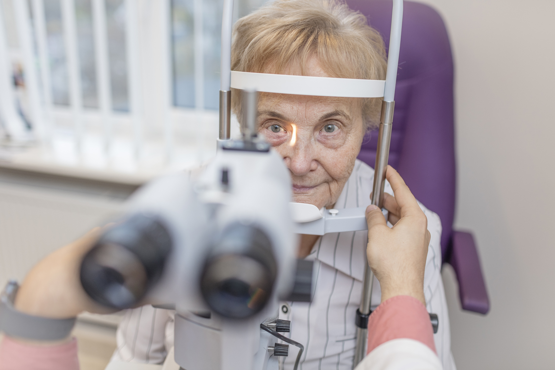
Is This the Next Best Thing to Regrowing a New Retina?
A BrightFocus Macular Degeneration Research-funded scientist is investigating a new regeneration technique to restore key cells that are wiped out in late-stage age-related macular degeneration.
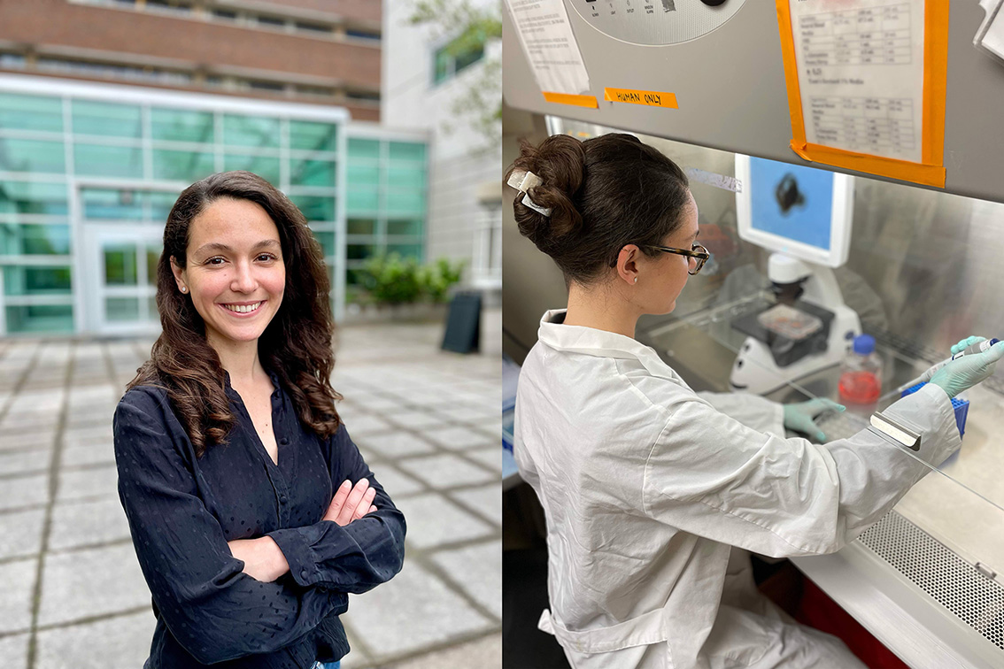
Scientists are looking to nature to guide new treatments. BrightFocus Macular Degeneration Research-funded researcher Juliette Wohlschlegel, PhD, explores whether the human retina can regenerate key vision cells damaged by macular degeneration.
The study focuses on coaxing Müller glia—support cells in the retina—into becoming cone cells, which are critical for seeing detail and color.
This regenerative approach could offer hope for restoring vision in people with late-stage age-related macular degeneration, when current treatments are no longer effective.
Just as a salamander can grow a new tail, the human retina may be capable of regenerating cells that allow us to see detail and color–If we can unlock potential with the right technique and ‘code’ for regeneration.
BrightFocus Macular Degeneration Research grant recipient Juliette Wohlschlegel, PhD, plans to harness the blueprints to regeneration found in nature. But rather than attempting to regrow the entire retina, she is working on a way to replace certain light-detecting retinal cells that are destroyed byage-related macular degeneration (AMD).
This novel approach could provide hope to people facing severe vision loss due to advanced AMD. “It offers the potential to restore vision in the later stages of the disease,” noted Dr. Wohlschlegel. Current treatments must be used much earlier, since they only stabilize eyesight in most people.
Borrowing from Nature to Fuel Human Discoveries
Juliette Wohlschlegel, PhD, Macular Degeneration Research grant recipient
Dr. Wohlschlegel is a neuroscientist with a deep drive to find potential treatments for people living with AMD. Her experience working in a geriatric hospital led her to pursue neuroscience as a student. She quickly became fascinated with vision research, a bond that grew even stronger after her own grandmother was diagnosed with AMD.
She expects her new study to reveal information that will bring the goal of developing better treatments for AMD one step closer. “I believe that findings from our work will pave the way to therapeutic strategies that can improve patients’ daily lives,” she said.
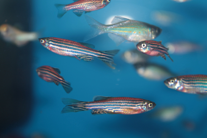
A handful of fish and amphibians have an amazing advantage over human beings and most other animals. If the retina in your eye is injured or damaged, a type of helper cell sets off a cycle of inflammation. This response typically leads to scarring and further damage. But a few species, including the small, striped zebrafish found in pet stores, respond to this type of trauma differently. They regrow a whole new retina. And those support cells—called Müller glia—play a key role in this renewal process.
In 2020, Dr. Wohlschlegel joined Dr. Thomas Reh’s lab at the University of Seattle in Washington. At the time, Dr. Reh (who is a leader in retinal regenerative medicine) and his team had already reached an important milestone toward the goal of restoring damaged retinal cells. They discovered that, under certain conditions, Müller glia in mice can transform into nerve cells or neurons.
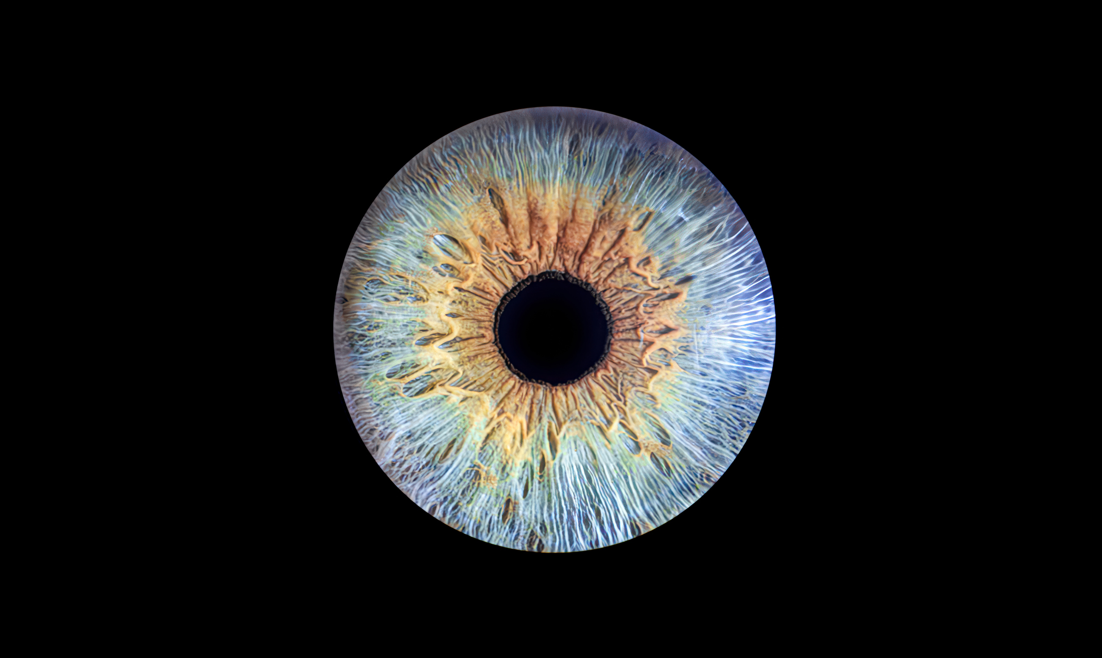
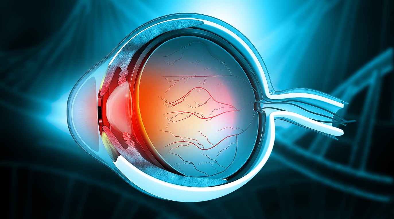
Light-sensitive nerve cells known as cones give us clear color vision. In AMD, cone cells in the macula die. The macula is the tiny ‘bullseye’ in the center of the retina with the greatest number of cones. By building on her own work and that of Dr. Reh’s team, Dr. Wohlschlegel hopes to coax Müller glia to turn into cone cells.
Dr. Wohlschlegel and her grandmother
Earlier research looking into why certain species can regrow the retina led to the discovery of a protein called Ascl1. Ascl1 allows immature neurons to develop into different types of mature, specialized nerve cells. When a zebrafish suffers injury or damage to the retina, the gene that makes Ascl1 goes into overdrive, unleashing a flood of the protein to kick-start the reprogramming. This repair process does not happen in mammals.
A Possible Path to Restoring Lost Vision
The next question was whether human retinal cells held the same potential to regenerate.
To find out, Dr. Wohlschlegel used human retinal cells taken from an organoid (a sort of mini-model of the retina). “We demonstrated that Müller glia derived from human retinal organoids can be reprogrammed into neurons,” Dr. Wohlschlegel said. But these new neurons, which were grown in a flat dish, did not keep their shape and stayed immature.
Dr. Wohlschlegel’s mentor, Dr. Thomas Reh
Next, Dr. Wohlschlegel’s team combined a method of growing cells in 3D with tools fine-tuned to target only Müller glia. This work showed “that Müller glia can be reprogrammed in 3D, and that the combination of factors influences the type of neurons the reprogrammed cells mature into,” Dr. Wohlschlegel explained.
Dr. Wohlschlegel’s current project is focused on finding a way to efficiently regenerate cone cells. “We will test different combinations of factors to promote cone production from human Müller glia,” she said.
Growing new cones from a person’s own Müller glia would avoid the drawbacks of more common approaches to replacing damaged retinal cells. Stem cell transplantation, for example, uses donor cells. To prevent the body from attacking the transplanted cells, people must take anti-rejection drugs. These medications make people more prone to serious infections and other health problems.
A Message From Dr. Wohlschlegel
Funding from Macular Degeneration Research has played an essential role in driving Dr. Wohlschlegel’s work forward. “It has had a significant impact on my research, enabling me to work more independently and concentrate on my own projects,” she said. Just as importantly, “it has provided me with the opportunity to join the vibrant community of BrightFocus researchers. I am truly honored and grateful to BrightFocus Foundation.”
About BrightFocus Foundation
BrightFocus Foundation is a premier global nonprofit funder of research to defeat Alzheimer’s, macular degeneration, and glaucoma. Since its inception more than 50 years ago, BrightFocus and its flagship research programs—Alzheimer’s Disease Research, Macular Degeneration Research, and National Glaucoma Research—has awarded more than $330 million in research grants to scientists around the world, catalyzing thousands of scientific breakthroughs, life-enhancing treatments, and diagnostic tools. We also share the latest research findings, expert information, and resources to empower the millions impacted by these devastating diseases. Learn more at brightfocus.org.
Disclaimer: The information provided here is a public service of BrightFocus Foundation and is not intended to constitute medical advice. Please consult your physician for personalized medical, dietary, and/or exercise advice. Any medications or supplements should only be taken under medical supervision. BrightFocus Foundation does not endorse any medical products or therapies.
Lessons from Zebrafish: Restoring "Ground Zero" in Macular Degeneration
BrightFocus Macular Degeneration Research grant recipient Lyndsay Leach, PhD, is investigating what makes another species capable of regrowing the part of the eye where age-related macular degeneration starts.
Article
How Understanding Lipid Processing in the Eye Could Spark Innovative AMD Treatment Approaches
With the help of funding from Macular Degeneration Research, Dr. Neetu Kushwah is studying the link between abnormal lipid regulation in the eye, inflammation, and age-related macular degeneration. Her findings could inspire new methods for treating the disease.
Article
New Supplement to Prevent Macular Degeneration Inspires Deeper Search for Cures
A Macular Degeneration Research-funded scientist launched a supplement based on a link he discovered between the eye disease and Parkinson’s disease. Now he’s continuing research in the hopes of finding novel cures.
Article
Transforming Macular Degeneration Care With Long-Acting Treatment
With funding from Macular Degeneration Research, Dr. Daisy Shu is exploring advanced drug delivery systems that release medicine slowly over time. This innovative approach could reduce the number of injections needed for macular degeneration while maintaining vision-saving results.
Article
How Targeting a Molecular 'Switch' Could Inspire New Macular Degeneration Treatments
A researcher funded by BrightFocus’ Macular Degeneration Research is zeroing in on a protein that damages eye cells in the early stages of dry age-related macular degeneration, offering insights that could lead to new treatments.
Article
How Better Models of Macular Degeneration Could Help Prevent Vision Loss
Most experimental models that researchers use to study macular degeneration fall short. BrightFocus Macular Degeneration Research grant recipient Dr. Brittany Carr aims to change that.