


Resources > Expert Information
National Glaucoma Research
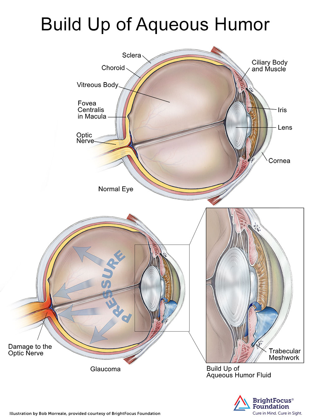
How the Build Up of Aqueous Humor Can Damage the Optic Nerve
Written By: BrightFocus Editorial Staff
Written By: BrightFocus Editorial Staff
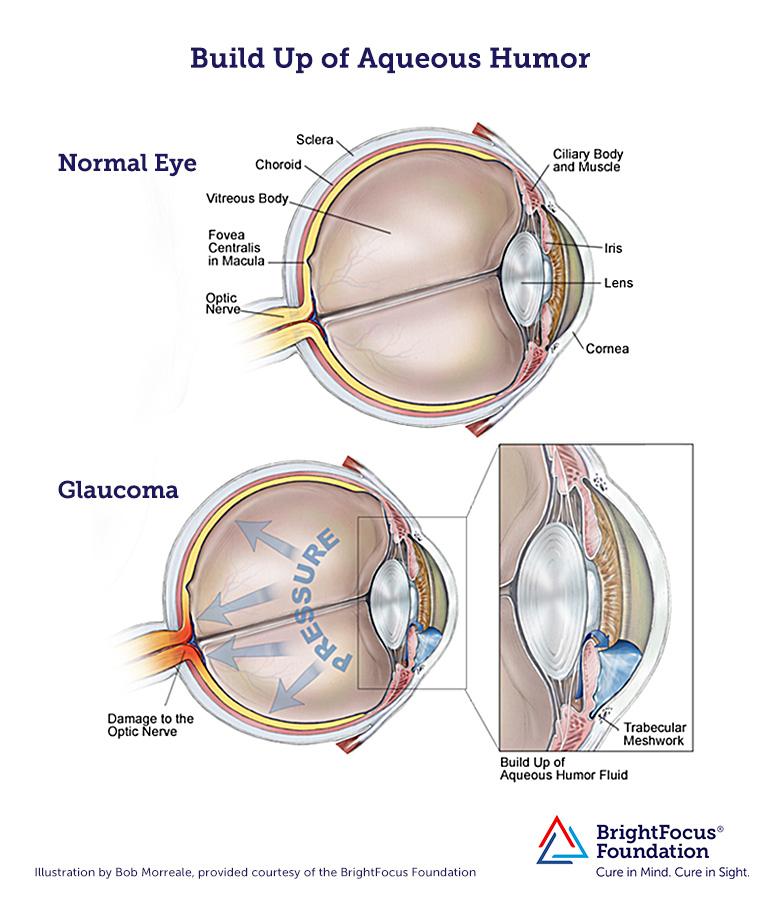 View a larger version of the medical illustration
Most, but not all, forms of glaucoma are characterized by high intraocular pressure. Intraocular pressure is maintained at normal levels when some of the fluid produced by the eye is allowed to flow out. The fluid (aqueous humor) is produced by the ciliary body where it flows into the anterior chamber and then out through a spongy tissue at the front of the eye called the trabecular meshwork into a drainage canal.
In open-angle glaucoma, fluid cannot flow effectively through the trabecular meshwork, and this causes an increase in intraocular pressure causing damage to the optic nerve and leading to vision loss.
Aqueous humor: Watery fluid that nourishes the interior of the front of the eye.
Ciliary body: Part of the eye, above the lens, that produces the aqueous humor.
Choroid: Layer of the eye behind the retina; contains blood vessels that nourish the retina.
Cornea: The outer, transparent structure at the front of the eye that covers the iris, pupil, and anterior chamber.
Fovea: The pit or depression at the center of the macula that provides the greatest visuals acuity.
Iris: The colored ring of tissue behind the cornea that regulates the amount of light entering the eye by adjusting the size of the pupil.
Lens: The transparent structure suspended behind the iris that helps to focus light on the retina.
Optic nerve: The bundle of nerve fibers at the back of the eye that carry visual messages from the retina to the brain.
Sclera: The tough outer coat that protects the entire eyeball.
Trabecular meshwork: Spongy tissue located near the cornea through which aqueous humor flows out of the eye.
Vitreous: Clear, jelly-like substance that fills the eye from the lens to the back of the eye.
BrightFocus Foundation is a premier global nonprofit funder of research to defeat Alzheimer’s, macular degeneration, and glaucoma. Since its inception more than 50 years ago, BrightFocus and its flagship research programs—Alzheimer’s Disease Research, Macular Degeneration Research, and National Glaucoma Research—has awarded more than $330 million in research grants to scientists around the world, catalyzing thousands of scientific breakthroughs, life-enhancing treatments, and diagnostic tools. We also share the latest research findings, expert information, and resources to empower the millions impacted by these devastating diseases. Learn more at brightfocus.org.
Disclaimer: The information provided here is a public service of BrightFocus Foundation and is not intended to constitute medical advice. Please consult your physician for personalized medical, dietary, and/or exercise advice. Any medications or supplements should only be taken under medical supervision. BrightFocus Foundation does not endorse any medical products or therapies.

Encontrará recursos e información que ofrecen productos, servicios y otro tipo de apoyo para personas con glaucoma y sus familias.
In clinical trials, stem cell therapy for glaucoma shows promise for rebuilding the eye’s drainage system and protecting the optic nerve.

Resources and information that provide products, services, and other support for people with glaucoma and their families.
Join us for a fascinating conversation with Dr. Lucy Q. Shen as we explore cutting-edge research into restoring vision loss from glaucoma.
Support Groundbreaking Glaucoma Research
Your support helps fund critical research that could prevent vision loss, provide valuable information to the public, and cure this sight-stealing disease.
Donate Today

